AI-generated summary by biochip.com, published . Not independently reviewed. Source: NASA Science, by Cinnamon Pritchard.
Key takeaways
- NASA says USB-drive-sized organ chips can model tissues such as lung, heart, pancreas, and liver.
- Chips can use adult volunteer cells from blood, skin, or stored cultures, then differentiate them into specialized tissue cells.
- The overview provides no clinical validation, accuracy data, or evidence that organ chips replace whole-body research.
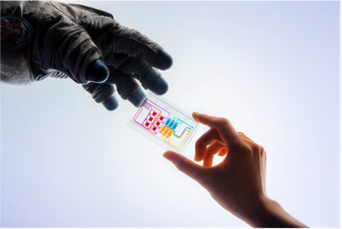
NASA Science describes organ chips as compact devices, sometimes about the size and shape of a USB drive, that let researchers study human tissues outside the body. Their central purpose is to show how tissues may respond to severe conditions, including radiation, and to treatments such as pharmaceuticals. Rather than relying on a single layer of cells in a dish, these chips contain cell cultures arranged to model the structures and functions of particular organs. NASA lists examples including the lung, heart, pancreas, and liver. Researchers can also connect individual tissue chips to imitate larger physiological systems, such as the blood circulatory system. A major practical feature is that the cells can come from adult human volunteers, obtained through a blood draw, skin cells, or stored cell cultures. Those starting cells can then be converted into the specialized cells needed for a chosen tissue model. The result is a small experimental platform intended to make studies of human biology, stress, and possible treatments more directly relevant to human tissues.
A Small Model of Human Tissue
An organ chip is not a miniature person or a fully formed organ. It is better understood as a carefully built test bed: a small device that carries living cells and is designed to reproduce selected features of a human tissue. Like a flight simulator that recreates the conditions a pilot needs to practice, an organ chip aims to recreate the tissue functions a researcher needs to observe.
The size matters because a device no bigger than a USB drive can make complex biology more manageable in the laboratory. NASA says these chips are used to investigate how human tissues respond to extreme stressors and therapeutic treatments. That focus places the chips between basic cell experiments and studies of whole organisms, offering a way to examine human cells in a defined experimental setting.
Modeling Specific Organs
The source identifies chips modeling the lungs, heart, pancreas, and liver. Each of these tissues performs different work in the body: lungs exchange gases, the heart drives circulation, the pancreas has key roles in metabolism, and the liver processes substances in the blood. A useful chip model therefore has to reflect the relevant structure and function of the tissue being studied.
That distinction is important because cells alone do not automatically behave like an organized human tissue. NASA describes organ chips as containing cell cultures that model both tissue structures and functions. In plain terms, researchers are not simply asking whether cells survive. They are trying to build a system in which the cells can be studied in an arrangement that better resembles their job in the body.
Connecting the Chips
Human organs do not operate as isolated parts, and NASA notes that organ chips can be linked to mimic entire physiological systems. The blood circulatory system is the example given in the source. Think of separate appliances joined by plumbing: each unit has its own task, but the connections determine how the larger system behaves.
For organ-chip research, linking models is a way to study relationships among tissues rather than viewing a lung, liver, or heart model entirely on its own. The source does not describe a particular linked-chip experiment or report performance results from one. Still, the ability to connect chips is central to the larger idea of representing coordinated human physiology in a compact laboratory platform.
Starting With Human Cells
NASA says organ chips can be made with adult human cells donated by volunteers. The starting material may come from a simple blood draw, from skin cells, or from cell cultures that have been stored. This gives the approach a direct human-cell foundation, rather than requiring the chip to begin with tissue from another species.
The collection routes also show why the platform can be flexible. A blood sample or skin cells are not themselves a lung, heart, pancreas, or liver tissue model. Instead, they provide cells that researchers can prepare and develop for the particular biological question they want to investigate.
How Cells Become Specialized
The source says cells can be differentiated, meaning they can be turned into specialized cell types for the tissues researchers want to study. Differentiation is the biological process by which cells take on more specific identities and roles. It is similar to training recruits for different jobs: they begin with shared potential, then receive the conditions needed to become specialists.
On an organ chip, this step makes it possible to move from donated or stored cells toward cells appropriate for a chosen tissue model. NASA does not specify the laboratory protocols, time required, or success rates for this conversion. It does establish the basic workflow: obtain human cells, generate specialized cells, and incorporate them into a device meant to model an organ or tissue.
Studying Stress and Treatments
NASA specifically highlights two broad uses: examining extreme stressors such as radiation and assessing therapeutic treatments such as pharmaceuticals. Radiation can place unusual stress on living tissue, while pharmaceuticals are substances intended to affect biological processes. Organ chips give researchers a controlled setting in which to observe how modeled human tissues respond to those exposures.
That controlled setting is valuable because researchers can focus on a selected tissue and a defined challenge. The source does not say that organ chips can replace all other forms of research, nor does it report that any treatment has been proven safe or effective through these devices. Their stated role is to support the study of tissue responses, not to provide a complete stand-in for the human body.
Why This Matters
Organ chips bring together several ideas that are useful for human health research: living human cells, models of specific organs, and the possibility of connecting tissue models into larger systems. For studies involving radiation or pharmaceuticals, that combination could help researchers ask more focused questions about how human tissues behave under stress or treatment. Their compact format also makes the technology distinct from studying an intact organ or person.
The most important caution is that a chip is a model. NASA's overview explains what these devices can be designed to represent, but it does not provide clinical outcomes, comparative accuracy data, or evidence that a particular chip predicts outcomes in patients. The value of any individual organ-chip study will depend on how well its cells, structure, and measured functions fit the question being asked.
What Comes Next
NASA's description points toward a research approach in which a volunteer's cells can be transformed into specialized tissue models and examined within a small device, alone or connected with other chips. Future work will need to show, for each use case, how closely a given model reflects the human tissue or system it is intended to mimic. As researchers apply these platforms to radiation, pharmaceuticals, and linked physiological systems, the key question will remain straightforward: what can this particular chip reliably tell us about human biology?