Source: American journal of obstetrics and gynecology, by Adi L Tarca; Roberto Romero; Sorin Draghici. AI-generated summary by biochip.com, published . Not independently reviewed.
DNA microarrays gave biology a way to read the activity of thousands of genes at once, turning what used to be a one-gene-at-a-time task into a broad survey of how cells behave. The source article explains how this approach is used for gene expression profiling, which means measuring how much messenger RNA, or mRNA, a cell is making from different genes. That matters because patterns of gene activity can reveal whether a disease is present, how aggressive it may be, and whether a patient is responding to treatment. In cancer especially, the article notes that microarrays can separate tumors that look similar under a microscope but behave very differently in the body. The piece also walks through the basic mechanics of the technology, from extracting mRNA to labeling it with fluorescent dyes and letting it bind to matching DNA probes on a chip. In simple terms, the method works a bit like laying thousands of tiny molecular fishing lines on a surface and seeing which genetic messages get caught. The article’s central point is that microarrays became an important discovery tool in clinical medicine because they connect lab measurements of gene activity to diagnosis, prognosis, therapy monitoring, and the search for disease mechanisms.
How a microarray reads gene activity
A microarray is essentially a dense grid of DNA probes attached to an inert surface. Each spot on that surface is designed to match a particular gene sequence, so when a sample is applied, the chip can test many genes in parallel rather than one by one.
The source compares this setup to a large parallel Southern or Northern blot, older lab methods used to detect DNA or RNA. The difference is scale: instead of separating material on a gel and probing a few targets, the microarray packs thousands of sequence-specific tests onto a single small platform.
From tissue sample to fluorescent signal
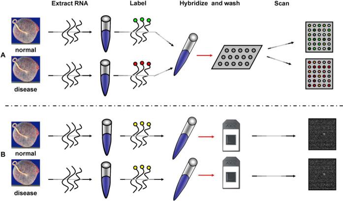
The process starts by extracting mRNA from tissues or cells. mRNA is the working copy a cell makes when it uses a gene, so its abundance serves as a proxy for how active that gene is.
Researchers then reverse-transcribe the mRNA into complementary DNA and label it with a fluorescent dye. That label acts like a visible tag, allowing instruments to detect where the sample binds after it is washed across the array.
Why matching sequences matters
The labeled sample is hybridized to the array, meaning the genetic material is given a chance to pair with complementary DNA probes fixed on the chip. You can think of it like matching pieces of Velcro: only sequences with the right molecular pattern should stick strongly to each other.
The article emphasizes that hybridization and wash steps are carried out under high stringency conditions. In plain language, that means the experiment is tuned to be picky, reducing cross-hybridization, where similar but not identical gene sequences might bind and create misleading signals.
Turning light into data
Once hybridization is complete, the array is scanned with laser-induced fluorescent imaging. The scanner reads the brightness at each sequence-specific spot and converts those light levels into numerical measurements.
The core assumption is straightforward: the amount of fluorescence at a given location is proportional to the amount of complementary mRNA in the original sample. Brighter spots suggest higher expression of that gene, while dimmer spots suggest lower expression.
What researchers do with expression profiles
The real power of microarrays comes from comparing expression patterns across samples. Instead of asking whether a single gene is on or off, researchers can look for broader signatures that distinguish healthy tissue from diseased tissue or one disease subtype from another.
According to the source, these comparisons can help identify diagnostic markers, which aid detection, and prognostic markers, which help estimate likely outcomes. They can also classify diseases such as tumors that may appear indistinguishable by routine microscopy but follow different clinical courses.
Applications in treatment and disease biology
The article also points to therapy monitoring as a major use case. If gene activity shifts after treatment, those changes may provide an early signal that a drug is working, failing, or triggering resistance.
Just as important, microarrays can help researchers understand disease mechanisms. When many genes move together in a pattern, that pattern can hint at the biological pathways driving the disease process, giving scientists clues about what is going wrong inside cells.
Limits hidden inside a powerful tool
Although the source highlights the promise of microarrays, its technical description also hints at why careful analysis is essential. Because the method depends on fluorescent intensity and sequence matching, small experimental errors in labeling, washing, or probe specificity can ripple into the final dataset.
That is why microarray studies usually require careful normalization and interpretation before any biological conclusion is trusted. Measuring thousands of genes at once is powerful, but it also increases the chances of noise, false patterns, and overinterpretation if the analysis is weak.
Why This Matters
Microarrays helped shift medicine toward a more data-rich view of disease. Instead of defining illness only by what tissue looks like under a microscope, clinicians and researchers could begin to describe it by the gene programs active inside cells.
That shift matters because diseases that share a name are often not biologically identical. A molecular profile can reveal hidden subtypes, suggest why one patient responds to therapy while another does not, and point researchers toward better targeted treatments.
The source presents microarrays as discovery tools, and that framing still captures their historical importance. Even as newer sequencing technologies have expanded what scientists can measure, the core idea behind microarrays endures: read many molecular signals at once, compare patterns carefully, and use those patterns to make sense of complex disease. The field has moved forward, but the logic described in this article remains foundational for modern biomarker research and precision medicine.