Source: Materials, by Yuanyuan Xu; Wenlong Yu; Yang Li; Lei Zhang (August 24, 2026). AI-generated summary by biochip.com, published . Not independently reviewed.
Key takeaways
- The review compares Transwell, microfluidic, tubular, self-assembled, and 3D-bioprinted blood-brain barrier model formats.
- Tsinghua University and Zhengzhou University researchers propose matched controls and minimum reporting for chemistry, mechanics, transport, and processing.
- The review calls for standardized comparisons but does not establish one best biomaterial or chip architecture.
Researchers at Tsinghua University and Zhengzhou University have assembled a practical review of how biomaterials and fabrication choices shape in vitro models of the neurovascular unit, the network of blood vessels and neural cells that supports the brain. The review centers on the blood-brain barrier, a selective cellular boundary that protects the central nervous system while also blocking many potential medicines. Its main message is that material chemistry, device manufacturing, cell culture conditions, and barrier measurements need to be considered together rather than as separate design choices. This is especially important for organ-on-a-chip systems, small microfluidic devices that give cells controlled flow, geometry, and chemical conditions. The authors compare conventional membrane-based models with microfluidic, tubular, self-assembled, and three-dimensional bioprinted systems. They focus on how matrix stiffness, degradability, biological binding sites, and device-body materials can influence barrier maturation and experimental reproducibility. The review also calls for matched controls and more consistent reporting of material properties, processing history, cell sources, flow conditions, and transport measurements. That framework could help researchers build more comparable blood-brain barrier models for drug screening, disease studies, and patient-specific testing.
Why the Neurovascular Unit Is Difficult to Recreate
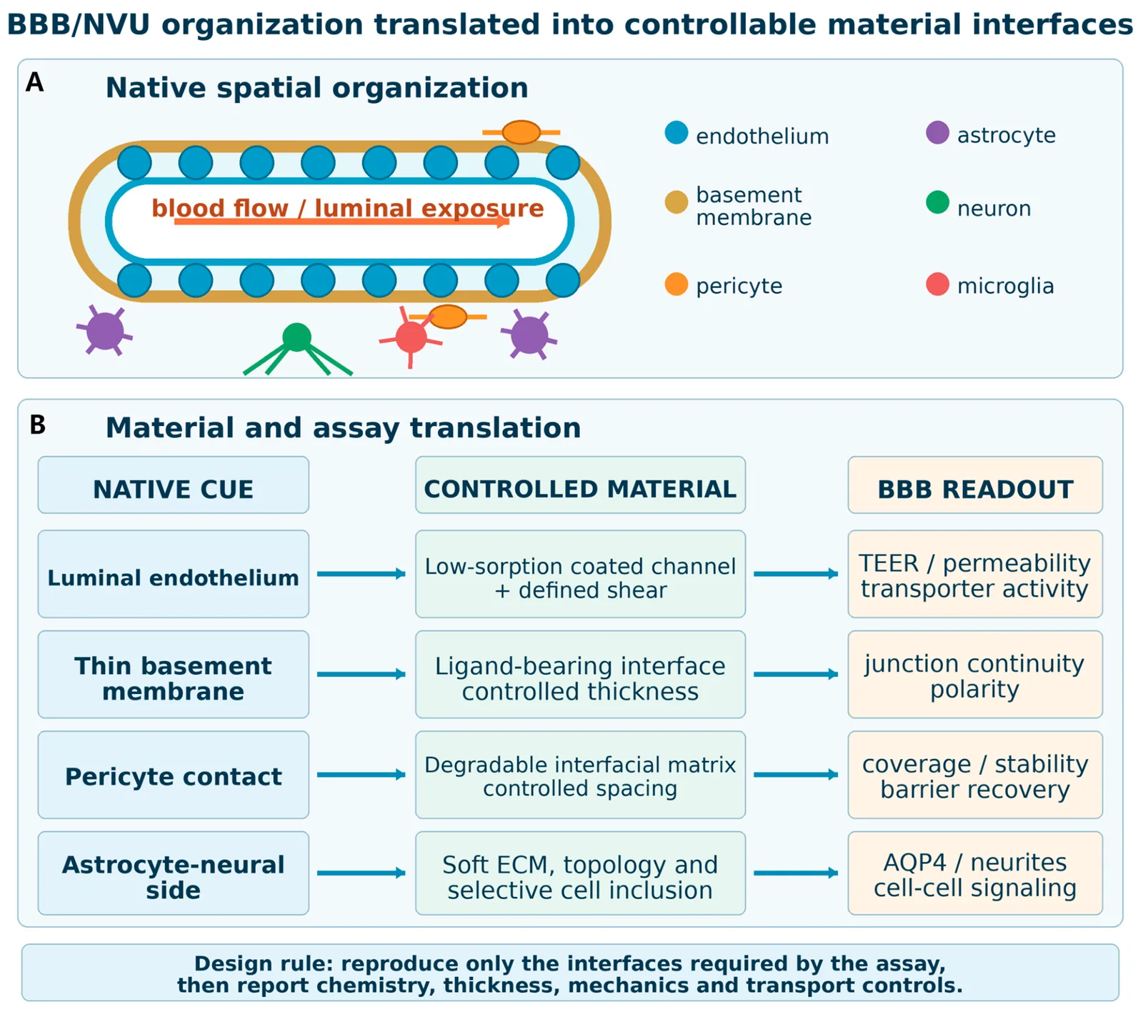
The neurovascular unit, or NVU, is the brain's tightly coordinated interface between circulating blood and nervous tissue. At its center is the blood-brain barrier, or BBB, where endothelial cells lining blood vessels form close junctions that carefully regulate what enters the brain. Basement-membrane components and nearby perivascular cells also help maintain that protective function.
Building an in vitro version is challenging because the BBB is not simply a layer of cells on a surface. It behaves more like a carefully staffed border crossing, where the physical barrier, chemical signals, cellular neighbors, and forces from flowing blood all affect which molecules can pass. A model that changes several of those factors at once may look sophisticated but still make it difficult to identify what caused a change in barrier performance.
Materials Are Part of the Experiment
The review organizes the field through a material-process-structure-function framework. In plain terms, the material selected for a model affects how it can be fabricated, fabrication determines the resulting physical structure, and that structure influences what cells do. This chain matters because cells sense their surroundings, including how stiff a matrix is, how quickly it degrades, and whether it presents molecules that cells can bind.
The authors examine natural, synthetic, semisynthetic, and decellularized extracellular-matrix hydrogels. An extracellular matrix is the supportive material around cells, similar to a building's structural framework and interior surfaces. Hydrogels can provide a water-rich environment that resembles aspects of native tissue, but their chemistry and preparation can vary substantially, affecting experiments that are meant to be compared.
Designing the Matrix and the Device Together
The review considers several ways to tune biomaterials, including crosslinking, peptide functionalization, stimuli-responsive materials, composite networks, and preparation methods. Crosslinking creates connections between polymer chains, much as ties can turn loose strands into a stronger net. Peptide functionalization adds short protein-like sequences that can give cells specific attachment cues.
These material decisions intersect with device engineering. Organ-on-a-chip platforms use microfluidics, the controlled movement of very small fluid volumes through channels, to expose cells to defined flow and biochemical gradients. The review links channel architecture, membranes or hydrogels, device-body materials, and fabrication routes to practical outcomes such as analytical access, reproducibility, and the development of a more mature barrier.
Comparing BBB Model Formats
Transwell systems, which typically separate cell compartments with a porous membrane, remain one format for studying barrier transport. Microfluidic systems add controlled flow and channel geometry, while tubular models aim to recreate vessel-like structures. Self-assembled and three-dimensional bioprinted models offer additional ways to arrange cells and matrices into more complex architectures.
No single format automatically captures every feature of the human BBB. The review instead emphasizes the tradeoffs among physiological complexity, the ability to measure transport, and the consistency needed for repeatable experiments. When researchers vary cell type, matrix chemistry, flow, device geometry, and barrier readouts simultaneously, it becomes hard to make head-to-head comparisons or derive design rules that transfer between laboratories.
Reporting Standards Could Make Models More Useful
The proposed solution is not a single preferred material or chip design. The authors call for matched controls and minimum reporting requirements covering chemistry, mechanics, transport, and processing. Reporting a hydrogel's composition alone is not enough when its stiffness, ligand density, degradation behavior, and preparation history may all alter how endothelial cells form junctions.
Consistent reporting could also clarify how cell source affects results. The review identifies induced pluripotent stem cell, or iPSC, and patient-derived cells as possible ingredients for individualized response testing. These approaches are promising because they could model disease-associated or patient-specific barrier dysfunction, but the material environment and device conditions must be sufficiently defined for results to be interpreted across studies.
Why This Matters
The BBB is a major obstacle in developing medicines for neurological disorders because it restricts the movement of many drugs into the central nervous system. It also changes in conditions such as brain tumors, where the blood-tumor barrier can become more permeable while retaining selective control over drug entry. Better in vitro models could complement animal studies, whose low throughput and species differences can limit translation to human drug research.
For biochip research, the key point is that a neurovascular chip is not merely a miniature container for cells. Its materials, interfaces, channel design, flow conditions, and measurement strategy collectively shape the biological behavior being measured. Treating those elements as an integrated system may make BBB platforms more useful for permeability and efficacy screening.
What Comes Next
The next generation of neurovascular models will depend on experiments that isolate individual design variables while retaining enough biological complexity to represent the BBB. Shared reporting practices could allow laboratories to compare matrices, fabrication methods, and chip architectures on more equal terms. That would give researchers a firmer basis for choosing models suited to questions about drug transport, barrier dysfunction, and individualized therapeutic responses.