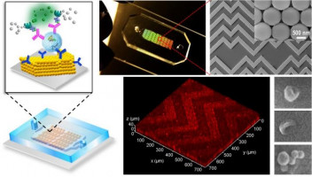
A new lab-on-a-chip device could make one of cancer medicine’s most promising ideas—liquid biopsy—faster, cheaper, and more sensitive. Instead of hunting for a tumor through surgery or imaging, liquid biopsy looks for biological clues circulating in the blood. In this case, the chip is designed to capture exosomes, tiny membrane-wrapped particles released by cells, including cancer cells, that carry proteins and genetic material. Researchers reported in Nature Biomedical Engineering that their device uses a clever 3D nanoscale design to pull these particles into close contact with a sensing surface, improving detection from very small blood samples. The work brings together engineering and cancer biology, with University of Kansas researchers testing the system on plasma from ovarian cancer patients. Their early results suggest the chip can spot signs of cancer in minuscule sample volumes, which could be valuable when sample collection is limited or repeated monitoring is needed. If the technology continues to perform well in larger studies, it could help move liquid biopsy from a specialized lab method toward a more practical clinical tool.
How the chip works
At its core, the device is a microfluidic chip, meaning it manipulates tiny amounts of liquid through channels smaller than a pinhead. These chips are attractive in medicine because they can run analyses quickly, use little material, and potentially lower costs.
The challenge is that exosomes are extremely small and hard to catch efficiently. Even when they flow near a sensor, a thin layer of liquid can keep them from making direct contact, reducing the odds that the chip’s molecular probes will grab them.
A nature-inspired design
The key innovation is a 3D nanoporous herringbone structure built into the chip’s surface. “Nanoporous” means it contains tiny holes at the nanometer scale, while “herringbone” refers to a zigzag pattern often seen in nature and in engineered surfaces that improve mixing.
That pattern helps stir the fluid and push exosomes toward the sensor surface, improving what engineers call mass transfer—the movement of particles from the flowing liquid to the place where they can be detected. Better mass transfer means more target particles reach the sensor in less time.
Why draining matters
According to researcher Zeng, previous microchannel designs had already found smart ways to improve mixing, but another problem remained. As particles approach a surface, the liquid trapped in the tiny gap between them creates hydrodynamic resistance, a force that acts like a cushion and makes direct contact harder.
The new chip solves that by letting the nanopores drain liquid from this gap. Zeng compared the idea to a sink full of water with floating balls on top: if you want the balls to touch the bottom, the easiest solution is to drain the water. In the same way, the chip removes enough liquid at the interface to bring exosomes into firmer contact with the sensing surface, where capture molecules can recognize them.
From engineering to patient samples
To build and test the device, Zeng collaborated with Andrew Godwin, a tumor biomarker expert and deputy director of the KU Cancer Center, along with graduate student Ashley Tetlow in Godwin’s Biomarker Discovery Lab. That kind of partnership matters because making a chip in the lab is only half the problem; proving it works on real patient material is the harder step.
The team evaluated the device using clinical plasma samples from ovarian cancer patients. Plasma is the liquid component of blood, and it can carry exosomes shed by tumors. The researchers found the chip could detect the presence of cancer using only a very small amount of plasma, a promising sign for future diagnostic use.
Why ovarian cancer is a compelling test case
Ovarian cancer is often difficult to catch early because symptoms can be vague and there is no simple, widely used screening test for the general population. That makes blood-based methods especially appealing, since they could offer a less invasive way to look for molecular signals of disease.
Exosomes are particularly interesting because they are not just debris. They carry biological information from tumor cells and may help cancers grow, communicate with surrounding tissue, and spread to other parts of the body, a process known as metastasis. Capturing them efficiently could therefore support both diagnosis and a better understanding of how disease is progressing.
Why This Matters
The broader significance of this work is not just that one chip performed well in one study. It points to a path for making liquid biopsies more practical by solving a basic physics problem: how to get rare nanoscale particles out of flowing blood samples and onto a sensor reliably.
If that can be done consistently, clinicians may be able to detect cancer-related signals earlier, monitor treatment response more often, and do so with smaller blood draws. A cheaper, more sensitive platform could also make advanced testing available beyond major research hospitals, which is often the real hurdle for promising medical technologies.
What comes next
The results are encouraging, but this is still an early-stage technology that will need larger clinical studies before it can become a routine diagnostic tool. Researchers will have to show that the chip works across more patients, different cancer stages, and potentially other tumor types, while also proving that results are robust in real-world laboratory settings.
Still, the concept is powerful: use nanoscale architecture not just to sense biology, but to physically guide it into contact with the sensor. If future trials confirm these early findings, this lab-on-a-chip could become part of a new generation of compact devices that bring sophisticated cancer testing closer to the point of care.