Researchers at the University of Kansas have developed a new lab-on-a-chip device designed to make liquid biopsy tests more sensitive. The chip focuses on detecting exosomes, tiny membrane-wrapped particles released by cells that can carry proteins and genetic material linked to cancer. Tumor cells shed exosomes into the bloodstream, which makes them a promising signal for spotting disease from a simple blood sample rather than an invasive tissue biopsy. In work reported in Nature Biomedical Engineering, the KU team showed that their microfluidic device could identify signs of ovarian cancer using only a very small amount of patient plasma. The advance comes from a new three-dimensional nanoporous design that improves how these nanoscale particles meet the chip’s sensing surface. In ordinary microfluidic systems, that contact step is a major bottleneck, because particles can hover near a sensor without actually touching it. By solving that transport problem, the KU researchers may have opened the door to faster and more reliable cancer screening tools built on compact chips.
A Better Way to Read Tiny Cancer Signals
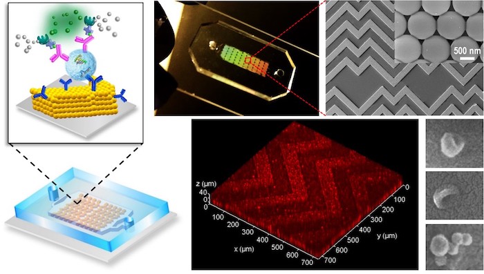
The project was led by researcher Steven Zeng, who described the challenge with a vivid analogy: imagine a sink full of water with lots of balls floating on the surface. If you want those balls to reach the bottom where sensors are waiting, the simplest trick is to drain the water. That basic idea shaped the chip’s design.
Exosomes are extremely small, far smaller than whole cells, and that makes them difficult to capture efficiently. Yet they matter because they can reflect what a tumor is doing, including signals tied to growth and metastasis, the process by which cancer spreads to other parts of the body. A device that can reliably isolate and analyze them could turn a blood draw into a much richer diagnostic test.
What Makes This Chip Different
The key innovation is a 3D nanoporous herringbone structure. “Nanoporous” means the material contains extremely tiny holes, while “herringbone” refers to a repeating V-shaped pattern often seen in natural structures and used in engineering to stir fluids. Together, those features help move exosomes toward the sensing surface much more effectively than standard flat designs.
That improvement addresses a problem known as mass transfer, the movement of particles from flowing liquid to a surface where they can be detected. In small channels, researchers can use clever patterns to nudge particles closer to the surface, but a thin layer of liquid still tends to remain between the particle and the sensor. That layer creates hydrodynamic resistance, essentially a fluid barrier that prevents efficient contact.
Zeng said the new structure drains liquid from that tiny gap, allowing exosomes to come into hard contact with the sensor surface. Once that happens, molecular probes on the chip can recognize and capture the particles. In plain terms, the device does not just bring biomarkers near the sensor; it helps them actually stick where they can be measured.
Built Through Collaboration
To develop and validate the chip, Zeng worked with Andrew Godwin, deputy director of the KU Cancer Center and a tumor-biomarker specialist in the Department of Pathology and Laboratory Medicine at the KU Medical Center. Graduate student Ashley Tetlow from Godwin’s Biomarker Discovery Lab also contributed to the effort. The collaboration brought together expertise in microengineering, cancer biology, and clinical sample analysis.
That mix matters because promising devices often fail when they leave the engineering bench and face real patient material. Blood plasma is a messy biological fluid full of molecules and particles that can interfere with measurement. Testing with clinical ovarian cancer samples gave the team an early indication that the chip could work under realistic conditions, not just in simplified lab experiments.
Why Ovarian Cancer Is a Strong Use Case
Ovarian cancer is especially difficult to detect early, and that is one reason liquid biopsy tools are attracting so much interest. Symptoms can be vague, and conventional diagnosis often happens after the disease has progressed. A sensitive test that reads tumor-derived exosomes from a small plasma sample could someday help clinicians spot disease earlier or track how it changes over time.
The KU study found the chip could detect the presence of cancer in a minuscule amount of plasma. That is important because lower sample requirements can make testing easier, less expensive, and more practical for repeated monitoring. In the long run, doctors want tools that can be used not only once, but many times during treatment to see whether a therapy is working or a cancer is returning.
Why This Matters
Lab-on-a-chip systems aim to miniaturize laboratory functions onto devices small enough to fit in the palm of a hand. If they work well, they can reduce the time, cost, and sample volume needed for sophisticated analyses. The challenge has been translating elegant chip designs into measurements sensitive enough for real disease detection.
This work matters because it tackles one of the core physics problems behind biosensing, not just the chemistry of recognizing a biomarker. By improving how nanoscale particles are delivered to the sensor surface, the KU team may have created a platform that could be adapted beyond ovarian cancer. Similar strategies could potentially be used for other tumor types or even for non-cancer conditions where extracellular vesicles and rare biomarkers carry useful information.
What Comes Next
The results are promising, but a research chip is not the same as a finished clinical product. Larger studies will be needed to confirm accuracy, determine how well the test performs across diverse patients, and compare it with existing diagnostic methods. Researchers will also need to show that the device can be manufactured consistently and used reliably outside specialized labs.
Even so, the study points to an important direction for precision medicine: better diagnostics may come not only from discovering new biomarkers, but from building smarter devices to find the ones already hiding in blood. If future trials bear out these early findings, nanoporous microfluidic chips like this one could help move liquid biopsy from a promising concept to a more routine part of cancer care.