A research team has developed a tiny microfluidic device, called an MPA-Chip, to pull circulating tumor cells or CTCs out of blood samples. CTCs are cancer cells that break away from a tumor and travel through the bloodstream, and doctors see them as a potential window into how a patient’s disease is progressing. The challenge is that these cells are extremely rare, so finding them reliably in a tube of blood is technically difficult. In this study, the researchers designed a chip filled with microscopic pillar-like structures and tested several pillar shapes to see which one best trapped tumor cells while letting most normal blood cells pass through. A lozenge-shaped design emerged as the top performer, combining strong capture rates, high purity, and excellent cell viability, meaning the trapped cells were still alive and potentially useful for later analysis. The chip was then tested on blood from 12 breast cancer patients, where it detected differences between non-metastatic and metastatic disease and even appeared to reflect response to chemotherapy in one patient over time. The work points toward a cheaper and more practical way to monitor cancer from blood rather than repeated tissue biopsies. If the approach continues to hold up in larger studies, it could help make cancer tracking more routine, faster, and less invasive.
A Small Chip for a Big Clinical Problem
CTCs have attracted intense interest because they offer a kind of “liquid biopsy,” a test that looks for cancer-related material in blood instead of surgically removing tissue. In principle, counting and studying these cells could help doctors judge prognosis, detect spread, and monitor whether treatment is working.
But there is a catch: CTCs are scarce. A blood sample contains huge numbers of red blood cells and white blood cells, while tumor cells may appear only in tiny numbers, so any useful device has to be both selective and gentle.
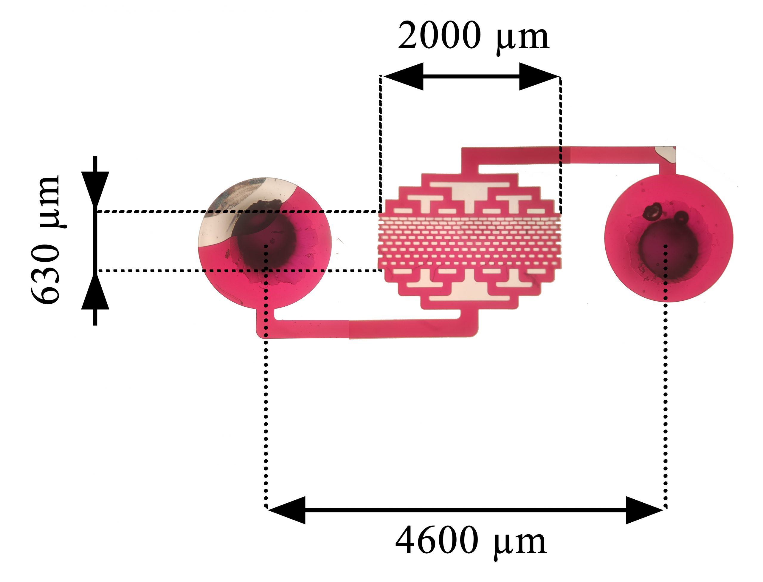
How the MPA-Chip Works
The new device is a micropillar array-based microfluidic chip, abbreviated as MPA-Chip. Microfluidics means controlling very small amounts of fluid through channels the size of a fraction of a millimeter, allowing researchers to sort or analyze cells with precision.
Inside the chip, blood flows through arrays of microscopic pillars. These pillars create physical conditions that make it easier to isolate tumor cells based on how they move, interact, and are retained within the chip, while most unwanted cells continue flowing through.
Why Shape Matters at the Microscale
The researchers did not settle on one pillar design from the start. They built and compared several geometries—lozenge, rectangle, circle, and triangle—and used numerical simulations to study how blood would move through each layout.
Those simulations examined features like velocity and pressure profiles, which describe how quickly fluid flows and how force is distributed inside the chip. At this scale, subtle changes in geometry can strongly affect whether fragile cells are captured efficiently or damaged in the process.
After the computer modeling, the team ran experiments using breast and prostate cancer cell lines as stand-ins for real tumor cells, along with blood samples. The goal was to measure three practical outcomes: capture efficiency, meaning how many target cells were trapped; purity, meaning how free the sample was from contaminating blood cells; and viability, meaning whether the captured cells remained alive.
The Lozenge Design Came Out on Top
Among the tested shapes, the lozenge geometry performed best. According to the study, the optimized lozenge MPA-Chip delivered capture efficiency above 85%, purity above 90%, and viability of 97%.
That combination matters because many devices do one thing well at the expense of another. A chip might trap many cells but collect too much cellular debris, or it might isolate cells cleanly but damage them so badly that downstream analysis becomes difficult.
The device itself was also compact. The MPA-Chip measured just 0.25 square centimeters, with a throughput of 0.5 milliliters per hour for a single chip, and the researchers noted that throughput could be scaled up by running multiple chips in parallel.
Testing the Chip in Breast Cancer Patients
To move beyond lab models, the researchers validated the chip using blood from 12 patients with breast cancer in different disease states. This is an important step, because real patient samples are much messier and more variable than cultured cells in controlled experiments.
The chip detected CTCs in both non-metastatic and metastatic patients. The reported median count was 6 CTCs for non-metastatic disease and 25 CTCs for metastatic disease, suggesting that the device may capture clinically meaningful differences tied to how far the cancer has progressed.
That is notable because metastasis—the spread of cancer to other parts of the body—is what makes many cancers far more dangerous. If a blood-based test can help distinguish lower-burden disease from more advanced disease, it may become useful as a monitoring tool alongside imaging and other clinical assessments.
A Glimpse of Treatment Monitoring
One of the most intriguing details in the report was its connection to chemotherapy monitoring. In a metastatic patient, the number of captured CTCs dropped from 23 to 7 over the chemotherapy period.
This does not prove on its own that the chip can serve as a definitive treatment-response test, especially given the small number of patients. Still, it hints at the real clinical promise of CTC analysis: a repeatable blood test that could show whether therapy is reducing the population of tumor cells circulating through the body.
The researchers also suggested that the platform could eventually do more than isolate and stain CTCs. Because the captured cells remained highly viable, future versions may support on-chip culture, which means keeping the cells alive for longer-term study and possibly testing how they respond to drugs.
Why This Matters
The importance of this work lies in its attempt to balance performance with practicality. Many CTC technologies are promising in theory but can be costly, complex, or difficult to translate into routine use, so a low-cost, efficient microfluidic platform would fill a real need.
If the MPA-Chip can be validated in larger and more diverse patient groups, it could support a less invasive way to follow cancer over time. Instead of relying only on scans or repeated tissue biopsies, clinicians might one day use blood samples to track disease status, estimate prognosis, and adjust treatment with better timing.
There is still a long road from a promising chip to standard clinical care. The study involved a small cohort, and broader testing will be needed to show how well the device works across cancer types, treatment settings, and real-world hospital workflows.
Even so, the results highlight how much impact careful engineering can have in medicine. By changing something as simple as the shape of microscopic pillars inside a chip, the researchers improved the capture of rare cancer cells and opened the door to richer information from an ordinary blood draw. As liquid biopsy technologies mature, devices like this could help turn cancer monitoring into something more frequent, less invasive, and more responsive to the realities of each patient’s disease.