AI-generated summary by biochip.com, published . Not independently reviewed. Source: Cancers, by Zeina Habli; Walid AlChamaa; Raya Saab; Humam Kadara; Massoud L Khraiche.
Circulating tumor cells, or CTCs, are cancer cells that break away from a tumor and travel through the bloodstream, and researchers see them as a kind of liquid biopsy that could reveal what a cancer is doing without a surgical tissue sample. The source article reviews how microfluidic chips—devices that guide tiny amounts of fluid through precisely engineered channels—are becoming central tools for finding and isolating these rare cells. That matters because CTCs are vanishingly uncommon compared with normal blood cells, so any useful test has to separate a few target cells from an overwhelming background. The review explains that newer chip designs combine biological recognition, such as antibodies that bind to tumor-cell markers, with physical sorting methods based on size, flow behavior, or magnetic labeling. Among the examples discussed are hybrid systems like the CTC-iChip, as well as antibody-based platforms such as the GEM chip, GEDI chip, LiquidBiopsy, and OncoCEE. Together, these devices aim to improve how sensitively and specifically clinicians can detect CTCs, while also preserving cells well enough for downstream analysis or even culture. The overall picture is hopeful but not simple: the field has real clinical promise, yet it still faces technical tradeoffs around sensitivity, selectivity, and the biological diversity of tumor cells.
Why CTCs are so hard to catch
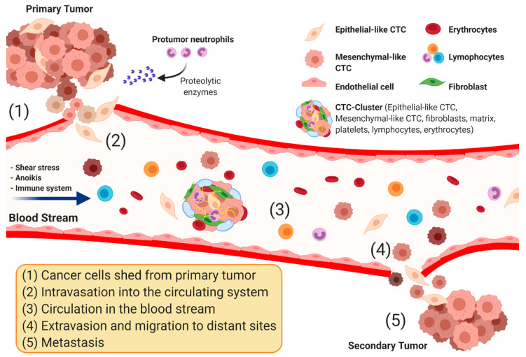
Looking for CTCs in blood is a bit like trying to pull a few specific grains of sand out of a swimming pool. The cells are rare, fragile, and mixed in with huge numbers of red blood cells, white blood cells, and platelets.
That rarity shapes the whole technology problem. A practical platform must enrich, or concentrate, tumor cells while washing away normal blood components without losing the very cells the test is meant to find.
How microfluidic chips help
Microfluidic chips tackle that problem by shrinking the assay onto a carefully fabricated device where fluid flow can be tightly controlled. In plain terms, they act like miniature plumbing systems that steer cells through narrow paths so that contact with chip surfaces happens in a predictable way.
The source emphasizes several advantages of this setup. Because the devices have controlled geometry, directed flow, and engineered surface coatings, they can increase the odds that a CTC will contact and stick to a capture surface while unwanted cells keep moving. Once target cells are immobilized on the chip, the remaining blood material can be flushed away, which improves enrichment against leukocytes, the white blood cells that often contaminate CTC samples.
Using antibodies as molecular hooks
Many CTC devices rely on antibodies, proteins that recognize specific markers on the cell surface. A useful analogy is Velcro: if the chip is coated with the right molecular hooks, cells carrying the matching surface marker are more likely to grab on as they pass.
The marker most commonly used is EpCAM, short for epithelial cell adhesion molecule, which is often found on tumor cells of epithelial origin. The review notes, however, that some platforms use a cocktail of antibodies tailored to a particular cancer type, while others work by negative depletion, using antibodies such as anti-CD45 or anti-CD66 to retain leukocytes and let CTCs pass through. That shift is important because not all tumor cells display the same markers, and some may lose EpCAM as they change state during metastasis.
Hybrid systems try to solve single-platform weaknesses
One of the review's central themes is that no single capture strategy is perfect. Marker-based methods can miss tumor cells that do not express the chosen antigen, while purely physical sorting methods may collect more unwanted cells along with the targets.
That is why a newer generation of platforms combines immunomagnetic beads with microfluidics. In these systems, tumor-associated cells are first labeled in whole blood with magnetic particles, then passed through a chip that further separates cells by size or flow behavior, including approaches such as inertial focusing. The idea is to pair the specificity of molecular labeling with the sorting power of microscale fluid control.
The CTC-iChip and other devices in the field
The review highlights the CTC-iChip as a promising example of this hybrid approach. According to the source, it integrates three microfluidic components in line within a single automated system, beginning with magnetic labeling of cells in whole blood and then sequential processing through multiple chip-based sorting steps.
The first component uses deterministic lateral displacement, a method that nudges cells into different paths based on size as they flow past an array of obstacles. After that, additional stages refine the separation so magnetically labeled cells can be isolated with greater sensitivity. The article also names several surface-based antibody capture systems, including the geometrically enhanced mixing or GEM chip, the geometrically enhanced differential immunocapture or GEDI chip, LiquidBiopsy from Cynvenio Biosystems, and OncoCEE from Biocept.
What these platforms could enable clinically
The practical appeal of CTC capture is not just counting cells. Once isolated, CTCs could be inspected under a microscope, profiled for molecular features, or potentially cultured for further testing, offering a repeatable blood-based window into a patient's disease.
The source argues that compared with standard immunomagnetic bead-only platforms, some microfluidic devices offer higher capture specificity and selectivity. In clinical terms, that could move CTC assays beyond prognosis—estimating how a disease may progress—toward diagnosis and treatment guidance. A cleaner, more intact CTC sample is more useful when doctors or researchers want to analyze mutations, protein expression, or changes over time.
Why This Matters
The bigger significance is that cancer care increasingly depends on measuring disease in real time, not just at the moment of diagnosis. Tissue biopsies can be invasive and difficult to repeat, while blood draws are routine, so a reliable CTC test could give clinicians a less burdensome way to track progression, relapse, and response to therapy.
At the same time, the review makes clear that the biology of CTCs is messy. Tumor cells are heterogeneous, meaning they vary from one another, and the very cells most relevant to metastasis may not fit neatly into one capture strategy. That is why engineering improvements alone are not enough; platform design has to account for the fact that cancer cells change their surface markers, size, and mechanical behavior as disease evolves.
The remaining challenges
Despite the progress, the field still faces a balancing act between sensitivity and purity. A system that captures more candidate CTCs may also pull in more background cells, while a highly selective system may miss important subpopulations.
Standardization is another hurdle. If different devices rely on different markers, different flow conditions, or different definitions of what counts as a CTC, it becomes harder to compare results across studies and translate them into routine clinical use. The review ultimately presents microfluidic CTC technology as a rapidly advancing toolkit rather than a finished solution, with the most promising future likely belonging to flexible, integrated platforms that can capture rare cells efficiently and preserve them for deeper biological analysis.