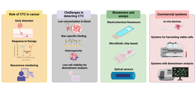
Circulating tumor cells, or CTCs, are cancer cells that break away from a tumor and drift through the bloodstream, and detecting them could give doctors a much earlier read on how a cancer is spreading or responding to treatment. The problem is scale: these cells are extraordinarily rare, often hidden among huge numbers of normal blood cells, which is why researchers often compare the task to finding a needle in a haystack. A recent wave of chip-based technologies is trying to solve that problem by turning a blood sample into a carefully controlled miniature lab. Instead of relying on one trick, these devices often combine several steps at once, such as sorting cells by size, capturing suspicious cells on specialized surfaces, and sometimes even keeping them alive for culture on the chip itself. The core idea is simple to picture: if blood is a crowded highway, microfluidic chips use tiny channels, obstacles, and chemical hooks to guide the vehicles into the right lanes. In the source material, researchers describe platforms that exploit differences in tumor-cell size, shape, deformability, and surface markers to enrich CTCs with much less sample loss than conventional handling. Two examples stand out: a multi-step microfluidic chip from Zhou and colleagues, and a biosensor-based detection platform from Burinaru and colleagues. Together, they show how biochips are moving CTC detection from a laborious lab procedure toward a more integrated, sensitive, and potentially clinically useful tool.
Why CTCs Are So Hard to Find
CTCs matter because they can carry real-time information about a patient’s disease without requiring a surgical biopsy. A simple blood draw, often called a liquid biopsy, could in principle reveal whether a tumor is shedding cells, changing behavior, or resisting therapy.
But blood is an unforgiving sample. CTCs are rare, while red blood cells, white blood cells, and other components vastly outnumber them, so even a small amount of cell loss during preparation can wipe out the signal researchers want to measure.
How Biochips Change the Search
Chip-based systems attack the problem by shrinking the workflow into tiny, precisely engineered channels. This field, known as microfluidics, manipulates small volumes of fluid so researchers can control how cells move, separate, and interact.
An everyday analogy is a well-designed airport security line. Instead of sending every passenger through the same path, the system uses lanes, barriers, and screening points to sort people quickly and consistently; a microfluidic chip does something similar for cells, using physical structures and surface chemistry to route rare tumor cells away from the crowd.
A Multi-Step Chip in One Device
Zhou and colleagues described a microfluidic chip that integrates size-based isolation, capture, and on-chip culture on a single platform. That matters because many older workflows involve multiple manual transfer steps between instruments, and each transfer creates a chance to lose the very cells researchers are trying to preserve.
The chip first uses a cell-separation channel to isolate larger CTCs from smaller blood cells through size-dependent migration. It then moves cells into a trapping chamber filled with diamond-shaped microposts, which retain CTCs based not just on size but also on deformability, meaning how easily a cell can squeeze and change shape under pressure.
This design reportedly achieved a separation efficiency above 94% for cells larger than 15 micrometers. The source also notes 97.4% cell viability during on-chip culture, suggesting that the platform does more than catch cells; it can keep them alive long enough for follow-up study.
Why On-Chip Culture Matters
Keeping captured CTCs alive opens a different level of analysis. Instead of merely counting cells, researchers may be able to observe how they grow, test drug responses, or study how aggressive they appear, all from a blood sample.
That is useful because cancer is not static. A tumor can evolve over time, and living CTCs may provide a window into those changes in a way that a one-time tissue biopsy cannot.
Biosensors Add a Second Layer of Detection
Some chip platforms go beyond physical sorting and add built-in sensors. Burinaru and colleagues developed a microfluidic biosensor for CTC detection based on electrochemical impedance spectroscopy, or EIS, a method that measures how electrical current is resisted as cells attach to a surface.
A simple analogy is stepping onto a smart floor that senses your presence by how your weight changes the signal. In the chip, when cells adhere to functionalized electrodes, they alter the electrical impedance, and that shift becomes a label-free way to detect captured cells without attaching fluorescent tags or dyes first.
How the EIS Platform Selects Cells
The Burinaru platform uses a PDMS, or polydimethylsiloxane, microfluidic chip integrated with interdigitated gold electrodes. These are comb-like electrode structures that increase the sensing area and make small electrical changes easier to detect.
The electrodes are coated with antibodies against EpCAM, a surface marker commonly found on many epithelial tumor cells, along with anti-CD36 antibodies to help improve selective capture while reducing non-specific interactions. The system then applies an alternating current, or AC, signal and reads the impedance changes produced by cell adhesion and interaction with the electrode surface.
This is important because physical enrichment alone is not always enough. A cell may be the right size yet still not be a tumor cell, so combining sorting with a molecularly targeted sensing step can improve specificity, the ability to identify the correct cells rather than just more cells.
The Design Logic Behind Better Detection
Across these examples, the bigger trend is integration. Researchers are combining filtration, selective capture, sensing, and sometimes cell culture into compact devices that reduce handling, shorten workflows, and preserve fragile rare cells.
That integration is especially valuable in CTC work because every extra wash, transfer, or labeling step introduces noise and loss. When the target is already vanishingly scarce, efficiency is not a minor engineering detail; it is the difference between seeing a meaningful biological signal and missing it entirely.
Why This Matters
If these chip-based systems continue to improve, they could make CTC analysis more practical for routine cancer monitoring. A clinician might one day use a blood sample to track whether a treatment is working, watch for early signs of metastasis, or collect living tumor cells for deeper analysis without an invasive procedure.
The source does not claim that these tools have solved every challenge. CTCs are biologically diverse, not all tumors shed them in the same way, and no single capture strategy will detect every cell type. Still, the progress described here shows that better fluid control, carefully designed microstructures, and integrated biosensors can substantially improve the odds of finding these rare cells. The next step is likely to be broader validation in real patient samples and tighter links between chip performance and clinical decisions, which is where a clever laboratory device becomes a useful medical test.