Source: Exploration of Targeted Anti-tumor Therapy, by Zhuldyz Myrkhiyeva; Kuanysh Seitkamal; Zhannat Ashikbayeva; Assiya Taizhanova; Daniele Tosi; Aliya Bekmurzayeva (May 29, 2025). AI-generated summary by biochip.com, published . Not independently reviewed.
Key takeaways
- Nazarbayev University researchers reviewed chip-based assays and biosensors for isolating and detecting circulating tumor cells.
- CTCs are rare blood-borne cancer cells linked to metastasis and potential liquid-biopsy applications.
- The review does not establish one universally validated platform for broad routine clinical use.
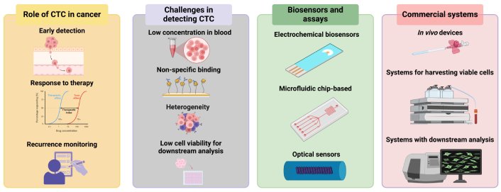
Researchers at Nazarbayev University have published a 2025 review of the tools used to find circulating tumor cells, or CTCs, the rare cancer cells that leave a primary or metastatic tumor and enter the bloodstream. These cells matter because they can travel to distant organs and contribute to metastasis, the spread of cancer that accounts for about 90% of cancer deaths. CTCs are also a potential component of a liquid biopsy, a blood-based test intended to provide information about a tumor without taking tissue directly from it. The opportunity is clear: a routine blood draw could potentially support early diagnosis, track response to treatment, and help forecast recurrence. The obstacle is equally clear, because CTCs exist at very low concentrations among a huge population of normal blood cells. Zhuldyz Myrkhiyeva, Kuanysh Seitkamal, Zhannat Ashikbayeva, Assiya Taizhanova, Daniele Tosi, and Aliya Bekmurzayeva examine chip-based assays, biosensors, and commercial detection platforms designed to address that problem. Their review frames CTC detection as a technical challenge involving the biology of the tumor cells, the complexity of blood, and the sensitivity of the measurement technology.
Finding a Rare Cell in Blood
CTCs are cancer cells that detach from tumors and enter the circulation through circulatory or lymphatic systems. Their connection to metastasis makes them especially important: cells moving through blood can reach secondary organs, where metastases may form. Thomas Ashworth first identified cells resembling tumor cells in the blood of a patient with metastatic cancer in 1869, establishing a concept that has become central to modern liquid-biopsy research.
The basic challenge resembles trying to find a particular grain of sand in a crowded beach. Blood contains a vast population of hematopoietic cells, the cells that make up the blood and immune system, while CTCs may be present at very low concentrations. A detection system must distinguish rare target cells without losing them during sample preparation or confusing them with the much larger background population.
Why CTCs Could Add Clinical Information
Unlike a conventional tissue biopsy, a CTC test begins with a blood sample. That simpler collection process could make repeated sampling more practical, allowing clinicians to monitor a patient over time rather than relying only on a single tissue specimen. The review identifies possible applications in early cancer diagnosis, monitoring response to therapy, and predicting disease recurrence.
CTCs are one element of liquid biopsy alongside circulating nucleic acids, including DNA, RNA, and microRNA, as well as exosomes and other tumor-associated cells. Their distinguishing feature is that they are intact tumor cells rather than fragments or molecules released into blood. Research summarized by the Nazarbayev University team indicates that CTCs may help predict disease progression and survival in metastatic cancer, and may also hold potential value in early-stage disease.
Three Layers of Detection Difficulty
The first problem is the cells themselves. CTCs are heterogeneous, meaning they can differ from one another in ways that affect whether a given assay recognizes or captures them. A method designed around one cellular feature may not identify every CTC in a patient's blood, particularly when tumor cells vary across primary and metastatic sites.
The second problem is the sample. Blood is not an empty transport fluid but a dense, complicated mixture of cells and biological material. The third problem lies in assay performance: detection platforms need enough sensitivity to identify rare cells while maintaining meaningful separation from non-target cells. Together, these factors can make CTC isolation and analysis technically demanding even when the clinical rationale is compelling.
Chip-Based Assays and Biosensors
Chip-based assays and biosensors are central to efforts to improve CTC detection. In plain terms, a biosensor is a device that turns a biological interaction into a measurable signal, while a chip-based assay places sample-handling and detection functions into a compact platform. For CTC work, these systems aim to help isolate rare tumor cells from blood and identify them with greater sensitivity.
Such platforms matter because detection is not a single step. A useful system must handle a complex blood sample, enrich or isolate the relevant cells, and produce a result that can be interpreted reliably. The review surveys methods developed for CTC isolation, including chip-based approaches and biosensors, while focusing on attempts to overcome low cell concentration, cellular heterogeneity, and limitations in measuring-platform sensitivity.
Commercial Platforms Face a Translation Gap
The team also considers commercially available platforms for CTC detection and the barriers to their broad clinical use. A commercial system may make CTC testing more accessible, but widespread adoption depends on more than the ability to detect a cell. The platform must perform consistently in the real-world setting where blood samples, tumor types, and CTC characteristics can vary.
The review does not present one platform as a universal answer. Instead, it emphasizes the unresolved hurdles that stand between promising detection methods and routine clinical application. That distinction is important: an assay can be technically capable of detecting CTCs while still facing questions about sensitivity, reliability across diverse cancer cases, and its role in treatment decisions.
Why This Matters
CTCs offer a compelling biological window into cancer because they are linked to the process by which tumors spread. If detection technologies can reliably retrieve and analyze these rare cells, clinicians may gain a way to repeatedly examine tumor-related information through a blood draw. That could complement tissue biopsy and other liquid-biopsy components rather than replace them outright.
The next advances will depend on platforms that can manage blood's complexity while recognizing the diversity of CTCs. By bringing chip-based assays, biosensors, and commercial systems into the same discussion, the Nazarbayev University researchers highlight a practical direction for the field: detection tools must be designed around both the rarity of the target and the clinical need for dependable results.
Topics: Biosensors · Cancer
Start here: Biosensors explained
Related articles
- Engineered Allosteric Biosensor for In Situ DNA Glycosylase Detection in Neuroblastoma Risk Stratification and Drug Resistance Monitoring
- Microfluidic biosensors for biomarker detection in body fluids: a key approach for early cancer diagnosis
- Companion Diagnostics in Clinical Therapy: Current Applications and Future Directions
- AI-Enhanced Photonic Biosensor Could Help Detect Glioblastoma Tissue