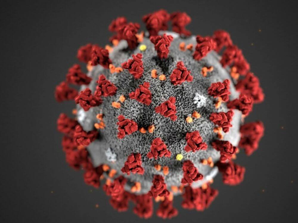
Microfluidics—the science of moving tiny amounts of liquid through channels thinner than a strand of hair—has emerged as a useful tool for studying and diagnosing SARS-CoV-2, the coronavirus that causes COVID-19. The core idea is simple: when a test only needs a droplet instead of a tube full of sample, it can often run faster, use fewer reagents, and fit into smaller devices. In the source material from Elveflow, microfluidics is presented not just as a way to speed up classic biochemical tests, but also as a platform for rebuilding aspects of human tissue in the lab. That matters because the virus does not infect isolated cells in a vacuum; it interacts with layers of tissue, fluid flow, and chemical signals that shape how infection begins and spreads. By recreating some of that complexity on a chip, researchers can study how the virus enters cells, how infection persists, and how drugs might interrupt those steps. The same miniaturized systems can also support diagnostics by making established lab methods more efficient and less sample-hungry. In short, microfluidics sits at the intersection of two urgent needs during a pandemic: better models of disease and faster, more practical testing. The article frames this technology as a bridge between basic virology, tissue engineering, and point-of-care diagnostics.
How microfluidics changes virus testing
Traditional lab tests for viral infection often depend on multiple preparation steps, specialized equipment, and a meaningful amount of sample. Microfluidic devices shrink those steps into compact pathways that can precisely guide fluids, a bit like replacing a bucket brigade with a network of narrow pipes and valves.
That miniaturization can make classic biochemistry-based tests faster and reduce the amount of sample required, according to Elveflow. For respiratory viruses such as SARS-CoV-2, that is especially appealing because testing demand can rise quickly and supplies can become constrained.
Why tiny channels can do big jobs
The power of microfluidics comes from control. At very small scales, engineers can regulate how quickly fluids move, how they mix, and when they meet cells or chemical reagents, giving researchers a more repeatable environment than many bulk experiments.
This matters for diagnostics because viral detection often depends on timing and contact between the sample and the sensing chemistry. If those interactions happen in a tightly controlled chip instead of a larger, less predictable container, the test can become more efficient and potentially easier to standardize.
Building infection models on a chip
The source also emphasizes a second role for microfluidics: creating better lab models of infection. Using tissue engineering, researchers try to build living systems that imitate features of human organs, and organ-on-chip devices push that idea further by placing cells in microengineered environments that mimic real tissues.
A helpful analogy is a movie set versus a real neighborhood. A flat layer of cells in a dish can show one important piece of biology, but an organ-on-chip adds structure, fluid flow, and multiple cell types, making the scene more realistic for studying how a virus behaves inside the body.
Recreating the cell's real environment
One of the main challenges in coronavirus research, as described in the source, is recreating the full cellular microenvironment. That means not only the cells themselves, but also the biological, chemical, and physical cues around them—the signals and forces that help determine whether a virus can attach, enter, replicate, and spread.
Microfluidic systems are well suited to this job because they can support 3-D architecture, multicellular complexity, and physiologically relevant biochemical forces. Those phrases sound technical, but the idea is straightforward: cells behave differently when they are arranged in lifelike structures and exposed to the kinds of flows and signals they would experience inside an airway or other tissue.
What this could reveal about SARS-CoV-2
Elveflow argues that microfluidic tissue-engineering could help researchers understand the coronavirus' entry strategy and the way infection persists in human cells. That is a practical research goal, because knowing exactly how the virus gains access and maintains itself can point to weak spots for treatment.
Microfluidic devices have already been used successfully for cell-based virus assays, according to the source. A cell-based assay is essentially a test that watches what happens when living cells encounter a virus, allowing scientists to measure infection, damage, or the effect of a candidate drug in a controlled setup.
From research platform to diagnostic tool
Although the article discusses broad COVID-19 background, its most concrete takeaway is that microfluidics can make diagnosis more compact and efficient. Coronavirus infections are diagnosed using features such as the virus's genomic sequence, its morphology, and disease-specific symptoms, and microfluidics can be integrated into testing workflows built around those biological signals.
The attraction is not that the chip replaces biology, but that it handles biology more neatly. Smaller volumes mean less waste, and integrated channels mean steps that might once have been spread across benchtop instruments can potentially be pulled together into one device.
Why This Matters
Pandemics expose two weaknesses at once: we often lack fast enough diagnostics, and we also lack realistic lab models for understanding a new pathogen. Microfluidics addresses both problems by shrinking tests for speed and by making disease models more lifelike than simple cell cultures.
That combination could shorten the path from basic research to practical response. If scientists can model infection more accurately and test samples more efficiently, they may be able to evaluate antiviral strategies sooner and improve access to testing in settings where sample volume, time, or equipment are limiting factors.
What comes next
The source stops short of presenting a single new clinical product or a specific trial result, but it lays out a clear direction for the field. As organ-on-chip systems improve and diagnostic chips become easier to manufacture, microfluidics could play a larger role in both outbreak preparedness and routine infectious disease care.
For SARS-CoV-2 and future respiratory threats, the broader lesson is that better tools often come from better scale. When researchers can manipulate fluids, cells, and signals in tiny, precise spaces, they gain a practical way to study infection more realistically and to build tests that are faster, smaller, and potentially more accessible.