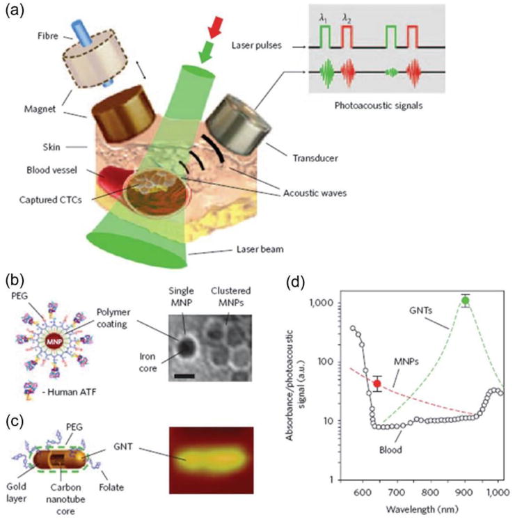
AI-generated summary by biochip.com, published . Not independently reviewed. Source: Chemical Society reviews, by Zheyu Shen; Aiguo Wu; Xiaoyuan Chen.
Circulating tumor cells, or CTCs, are cancer cells that break away from a tumor and travel through the bloodstream, and they have become an intense focus because a simple blood draw could one day reveal how a cancer is changing in real time. The source article reviews one of the most active areas in that effort: technologies that can find and isolate these extremely rare cells from a huge background of normal blood cells. Among the methods discussed, microfluidic chips stand out because they guide tiny amounts of fluid through narrow channels, almost like running a river through a carefully designed maze to trap only the cells researchers want. According to the review, these chips can work directly with peripheral blood and often avoid extra preparation steps such as dilution, labeling, or fixation. That matters because every extra handling step can lose cells or alter them before scientists have a chance to study them. At the same time, the article is clear that current systems still face practical barriers, especially when labs need to process larger blood volumes quickly and reliably. In short, the field has made real progress, but turning these devices into routine clinical tools will require better speed, scale, and consistency.
Why CTC detection is so hard
Detecting CTCs is difficult for a simple reason: they are rare. A tube of blood contains enormous numbers of red and white blood cells, while tumor cells may appear only a few times in each milliliter.
That is why most CTC technologies focus first on enrichment, which means pulling out likely tumor cells before trying to confirm exactly what they are. It is similar to panning for gold: before you inspect individual flakes, you need a way to separate them from everything else in the stream.
How microfluidic chips approach the problem
Microfluidics is the science of controlling fluids at very small scales, usually in channels smaller than a millimeter. The review notes that the field emerged in the early 1980s and later fed into technologies such as DNA chips, inkjet printheads, lab-on-a-chip systems, and micro-thermal devices.
For CTC detection, microfluidic chips use those tiny channels to manage how blood moves across surfaces or through structures designed to capture rare cells. In some designs, ligands—molecules that bind to specific targets—are attached to the chip so that tumor cells in flowing blood stick while healthy blood cells pass through.
What makes these chips attractive
The review highlights several practical advantages of microfluidic chip enrichment. One of the biggest is that patient blood can be applied directly to the chip without pre-dilution, pre-labeling, pre-fixation, or other processing steps.
That simplicity is useful because it can cut labor and reduce the chance of losing fragile cells before analysis begins. The authors also note that chip costs are not especially high, which is important if a technology is ever going to move from specialized research labs into wider clinical use.
The chips described in the review also show respectable sensitivity. The article reports that about 70 percent of CTCs can be captured when they are present at roughly 3 to 5 cells per milliliter of blood, a meaningful benchmark given how scarce these cells are.
Another major strength is what happens after capture. The enriched CTCs can be released from the chip for later phenotypic identification—looking at observable cell traits—and molecular analysis, which means studying DNA, RNA, or proteins to understand the biology of the cancer.
A closer look at the SIM-Chip system
The review points to a specific example from Kim and colleagues: a single-cell isolation platform called SIM-Chip. This system combines a microfluidic chip with immunomagnetic nanobeads, tiny magnetic particles coated so they can help bind and pull out target cells.
SIM-Chip is built as a two-step cascade platform. First, a lateral magnetophoretic microseparator enriches CTCs from whole blood using those immunomagnetic nanobeads; then a microdispenser handles the enriched cells one by one.
The second step is especially important because cancer researchers often want to study single cells rather than mixed populations. The platform electrically identifies cells using a single-cell impedance cytometer, which measures how a cell changes an electrical signal, and then isolates individual cells with what the paper calls a microshooter.
The source gives a few performance details that show how engineered these systems have become. The average droplet volume was 4.5 microliters, matching simulation results from an equivalent circuit model, and the microdispenser achieved a single-cell isolation throughput of 700 milliseconds, timed to buffer delivery and 96-well plate positioning.
The main bottlenecks
Even with these strengths, the review does not present microfluidic chips as a solved problem. One limitation is throughput: current chips struggle to handle large, milliliter-scale volumes of whole blood efficiently.
That may sound technical, but the implication is straightforward. If a clinic needs to screen blood quickly, a device that works beautifully on small amounts but slows down on larger samples may be hard to fit into routine care.
The review also points to flow rate as a challenge. Because microfluidic systems often run blood slowly through narrow channels to improve capture, the enrichment process can take a long time.
There is also a deeper tradeoff in many CTC platforms. The more carefully a chip tries to catch rare cells, the more it may sacrifice speed, and the more it tries to move fast, the more likely it may be to miss some cells or increase sample complexity downstream.
Why This Matters
Better CTC detection could make cancer monitoring less invasive. Instead of relying only on tissue biopsies, which can be painful and sample just one part of a tumor, clinicians could potentially use blood tests to watch how a disease evolves over time.
That could help with early assessment of treatment response, tracking metastasis, and studying how resistant cell populations emerge. The review makes clear that this promise depends not only on finding CTCs, but on isolating them intact enough for phenotype testing and molecular profiling afterward.
Microfluidic chips are appealing because they bring several needs together in one platform: direct use of blood, decent capture sensitivity, relatively low cost, and the possibility of releasing cells for deeper analysis. The remaining engineering problems—especially scaling to larger blood volumes and shortening processing time—are exactly the kind of issues that often determine whether a good lab method becomes a useful medical tool.
The broader takeaway is that CTC detection is moving from simple counting toward richer, cell-by-cell analysis. If researchers can keep improving throughput and reliability, microfluidic systems like SIM-Chip may help turn a rare cell in a blood sample into a practical source of information for cancer diagnosis, monitoring, and treatment decisions.