Source: Chembiochem, by Michael Chang; Lilly Nash; Ruben Bierings; Kei Takahashi‐Yamashiro; Takuya Terai; Tom Carter; Robert E Campbell; Kelvin K Tsao (August 26, 2026). AI-generated summary by biochip.com, published . Not independently reviewed.
Key takeaways
- University of Tokyo-led researchers developed serapH, an mStayGold-based pH biosensor bright at pH 7.4 and dim at pH 5.5.
- A carbon dioxide agar-colony screen measured fluorescence and pH sensitivity together, increasing colonies screened per engineering round.
- The reported work does not establish clinical use or performance across all live-cell imaging settings.
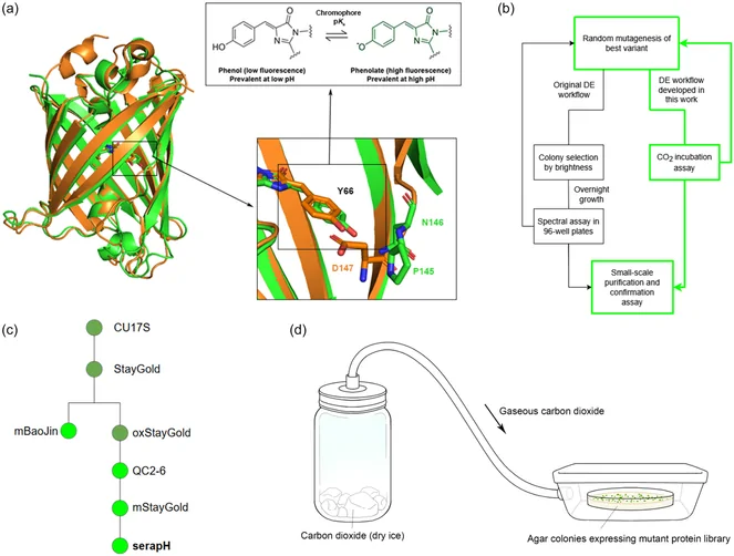
A team led by researchers at the University of Tokyo has developed serapH, a genetically encoded fluorescent biosensor designed to report changes in pH while resisting the fading that limits many existing probes. The sensor uses mStayGold, a fluorescent protein chosen as the engineering scaffold, rather than the more familiar green fluorescent protein from Aequorea victoria. In its reported form, serapH1.0 is bright at the near-neutral pH of 7.4 and dim at the acidic pH of 5.5. That behavior could help researchers follow cellular structures whose internal acidity changes as they move or release their contents. The team also created a colony-based screening approach that measures both fluorescence brightness and pH sensitivity directly on bacteria growing on agar. By using carbon dioxide to alter conditions around the colonies, the method increased the number of variants that could be evaluated in each round of protein engineering. The work addresses a practical imaging problem: fluorescent pH sensors can be useful only as long as they remain bright enough under repeated illumination. SerapH's improved photostability could support longer observations and higher excitation intensities in studies of pH-linked processes such as exocytosis.
Why Cellular pH Needs a Better Reporter
pH is a measure of how acidic or alkaline an environment is. Cells carefully control pH inside different compartments, and those values can shift during important activities such as endocytosis, when cells bring material inward, and exocytosis, when they fuse a membrane-bound compartment with the cell surface to release material.
Secretory organelles are a useful example. Synaptic vesicles, which store chemical messengers in nerve cells, have an internal pH of about 5.5, while endothelial-cell Weibel-Palade bodies typically rest between pH 5 and 5.5. When such compartments fuse with the cell membrane, their contents encounter the more neutral environment outside the organelle.
From Fluorescence to a pH Readout
Fluorescent proteins act a little like molecular light bulbs: researchers illuminate them with a chosen wavelength, and the proteins emit light that can be recorded by a microscope. A pH-sensitive fluorescent protein changes its brightness as acidity changes, turning a chemical condition into an optical signal.
Earlier sensors derived from the green fluorescent protein of the jellyfish Aequorea victoria, including superecliptic pHluorin and Lime, have been widely used for this purpose. Their weakness is low photostability, meaning that repeated exposure to excitation light can cause their fluorescence to fade, a process often called photobleaching.
Engineering serapH From mStayGold
The researchers built serapH using mStayGold as a starting scaffold. A scaffold is the parent protein structure that scientists modify, much as an engineer may alter a durable base design while preserving the features that make it reliable.
The resulting sensor was designed through directed evolution, an iterative protein-engineering process in which many variants are made and screened for useful traits. The reported serapH1.0 variant is highly fluorescent at physiologically neutral pH 7.4 and substantially dimmer at acidic pH 5.5, giving it the contrast needed to distinguish those conditions.
A Faster Way to Screen Protein Variants
Finding a useful sensor requires evaluating large numbers of mutant fluorescent proteins. The team developed a screening method that directly assessed both brightness and pH responsiveness in bacterial colonies on agar, rather than treating those features as separate tests.
Carbon dioxide incubation enabled the colony-based screen to probe pH sensitivity while fluorescence measurements identified bright variants. This approach increased the number of colonies that could be screened in each engineering round and reduced the time needed for each round, two advantages when researchers must search through many candidate mutations.
What Photostability Changes
Photostability matters because live-cell imaging often involves taking many images over time. A sensor that fades quickly can make a real biological change look smaller than it is, or force researchers to use gentler imaging conditions that miss fast events.
SerapH's improved resistance to fading compared with GFP-based pH sensors should allow higher excitation intensities and longer imaging durations. In practical terms, that could improve spatiotemporal resolution, the ability to see where an event occurs and when it happens, during pH-sensitive cellular processes.
Why This Matters
The development links protein design to a concrete sensing challenge. Rather than simply making a brighter fluorescent protein, the researchers sought a probe that combines pH-dependent behavior with durability under illumination, qualities that are both necessary for observing acidic secretory compartments over time.
The work also highlights the value of the screening system itself. A method that evaluates brightness and pH sensitivity simultaneously can make directed evolution more efficient, helping researchers prioritize variants that retain visible fluorescence while responding to the chemical signal of interest.
Next Questions for Live-Cell Imaging
SerapH provides a photostable option for studying pH-associated biology, including vesicle fusion and secretion. Its eventual utility will depend on how it performs across imaging conditions and cellular settings, but the mStayGold-based design and agar-colony screen offer a defined route for refining fluorescent biosensors for dynamic cell biology.