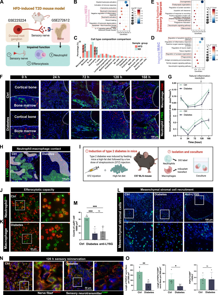
Researchers studying diabetic bone repair report that damaged sensory nerves may be a hidden reason these injuries heal so poorly. In their experiments, diabetic bone defects showed a double problem: neutrophils, the immune system's fast first responders, arrived late and then lingered too long. That combination can keep inflammation stuck in the wrong phase, which is especially harmful in bone healing, where cleanup must give way to rebuilding. The team designed a biomaterial scaffold to address both issues at once, using a fast pulse of interleukin-8 to draw neutrophils in early and a slower release of nerve growth factor to encourage sensory nerves to grow back. In diabetic rats, this two-step strategy improved mandibular bone regeneration by reshaping how nerves and immune cells interacted inside the defect. The work also points to a specific mechanism: regrowing neurites, or young nerve extensions, appeared to help macrophages recover their ability to clear aging neutrophils through a process called efferocytosis, partly through neuronal Galectin-3. Taken together, the study suggests that fixing inflammation in diabetes may require more than controlling immune cells alone; it may also mean restoring the sensory wiring that helps direct them.
A bone-healing problem with an immune and nerve component
Diabetic bone defects are notoriously hard to treat, and the paper argues that uncontrolled inflammation is a central reason. In healthy healing, inflammation is tightly timed: neutrophils rush in to contain damage, macrophages clean up spent cells, and then tissue-building processes take over. In diabetes, that sequence can break down.
The researchers link that failure to diminished sensory innervation, meaning fewer or weaker sensory nerve fibers in the injured tissue. Peripheral neuropathy, nerve damage outside the brain and spinal cord, is common in diabetes, but its role in local immune control has remained unclear. This study places sensory dysfunction much closer to the center of the story.
What went wrong inside diabetic defects
Inside diabetic bone defects, the team observed disrupted immune dynamics on two fronts. Neutrophil chemotaxis, the guided movement of these cells toward chemical distress signals, was delayed. At the same time, neutrophils were abnormally retained in the wound instead of being removed on schedule.
That retention mattered because old neutrophils are not meant to stay indefinitely. Normally, macrophages act like a cleanup crew, engulfing dying neutrophils in a noninflammatory process called efferocytosis. In the diabetic defects studied here, macrophage efferocytosis was impaired, allowing inflammation to smolder rather than resolve.
A scaffold built like a chocolate chip cookie
To intervene, the researchers created what they describe as a chocolate chip cookie-like scaffold. The image is useful: the embedded microspheres are the “chips,” and they release interleukin-8, or IL-8, in a burst. The surrounding silk fibroin matrix, the “cookie” around them, releases nerve growth factor more slowly over time.
This design was meant to control healing in sequence rather than all at once. IL-8 is a signaling protein that attracts neutrophils, so an early burst can help restore timely immune arrival. Nerve growth factor supports sensory nerve regrowth, which the authors propose is needed later to steer neutrophils toward a more pro-healing state and help macrophages resume proper cleanup.
Why timing mattered
The study found that timely neutrophil recruitment did more than restart inflammation. Early neutrophil chemotaxis induced by IL-8 also triggered bone healing through stem cell recruitment, linking the initial immune response to later regeneration. In other words, getting neutrophils to the site on time seemed to help set the stage for repair rather than just defense.
Sensory innervation then reinforced that effect by promoting neutrophil N2 polarization. Polarization is a shift in behavior, almost like changing a worker's assignment on a job site. Here, the N2 state refers to a more healing-associated neutrophil program, suggesting that regrowing nerves did not simply sit beside the immune response but actively shaped it.
A close partnership between nerves and macrophages
One of the paper's most interesting observations was physical. Macrophages preferentially positioned themselves close to outgrowing neurites and formed what the authors describe as a synapse-like structure. A synapse is normally thought of as a communication junction between nerve cells, so the comparison suggests a highly organized local interaction rather than random contact.
At those interfaces, the study found evidence that neurons helped restore macrophage efferocytosis through Galectin-3, a protein involved in cell signaling and adhesion. A simple way to think about it is that the nerves may be supplying missing instructions that tell macrophages when and how to finish the cleanup job. If that interpretation holds up, it adds a new layer to how diabetic inflammation might be corrected.
Testing the idea in diabetic rats
The researchers tested their spatiotemporal strategy in mandibular bone defects in diabetic rats. “Spatiotemporal” here means controlling both where signals are delivered and when they appear. That matters because wound healing is not just about which molecules are present, but about their order, location, and duration.
According to the study, regulating this neuroimmune circuit enhanced bone regeneration in the animals. The source summary does not provide detailed numerical outcomes, but it clearly frames the effect as improved mandibular repair compared with the uncontrolled diabetic condition. That makes the scaffold more than a conceptual tool; it functioned as a therapeutic system in a disease-relevant model.
Why This Matters
This work pushes diabetic bone healing beyond the usual idea that inflammation is simply too strong or too prolonged. It suggests that the real problem may be a broken conversation between sensory nerves and immune cells. If nerves help choreograph when neutrophils arrive, how they behave, and whether macrophages can clear them away, then neuropathy may directly sabotage healing at the injury site.
That has practical implications for biomaterials and regenerative medicine. Many scaffolds aim to deliver growth factors or recruit stem cells, but this study argues that successful repair in diabetes may depend on programming a sequence of immune and nerve events. Designing implants that restore those local instructions could become a more effective way to treat chronic defects.
What comes next
The findings open several next questions. Researchers will need to test whether the same neuroimmune mechanism appears in other bones, other diabetic injury models, and eventually in human tissue. They will also need to define how durable the regenerated nerve-immune interactions are and whether proteins such as Galectin-3 can be targeted directly.
Even so, the central message is already clear. In diabetic healing, the issue may not be just too little bone growth or too much inflammation, but poor coordination between systems that normally work together. By rebuilding that coordination with timed signals in a scaffold, the study offers a concrete path toward therapies that help chronic wounds and bone defects move forward again.