Biochip technology has moved far beyond tiny lab tools that simply hold samples in place. In the work summarized in this review, researchers show how these platforms have evolved into living, dynamic systems that can model human tissues such as skin and lung in ways standard cell culture often cannot. The big shift is from lab-on-a-chip devices, which miniaturize chemical and biological experiments, to organ-on-a-chip systems, which use microfluidics, living cells, and built-in sensors to imitate how real tissues behave. That matters because drug testing and disease research depend on models that are both realistic and practical. The review highlights skin-on-a-chip designs that can track barrier function, inflammation, and tissue metabolism in real time, as well as lung-on-a-chip platforms used to study viral infection and test antiviral compounds. These examples show a field trying to replace oversimplified petri-dish experiments with something closer to the body’s actual environment. The result is not just a better gadget, but a more useful bridge between basic biology, toxicology, and medicine.
From miniature labs to living tissue models
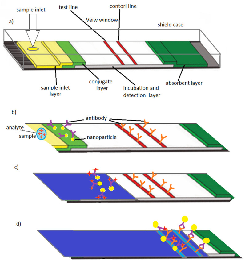
A useful way to think about a biochip is as a tiny workshop built onto a small device. Early lab-on-a-chip systems focused on shrinking lab procedures such as mixing fluids, detecting molecules, or separating samples into compact formats that used less reagent and ran faster.
Organ-on-a-chip, often shortened to OOC, adds another layer: living cells arranged in structures that mimic the architecture and flow conditions of real tissue. Instead of asking only whether a molecule is present, these systems can ask how cells respond over time, under stress, or during treatment.
Skin-on-a-chip gets closer to real tissue behavior
Skin is a strong candidate for organ-on-a-chip modeling because it is layered, exposed to the outside world, and central to drug and cosmetic testing. But ordinary cell culture struggles to capture how the epidermis, dermis, blood vessels, and immune cells interact inside actual skin.
The review describes a platform from Alexander and colleagues that integrates biosensors directly into a skin-on-a-chip system. That design measures transepithelial electrical resistance, a readout of how well a tissue barrier is holding together; in simple terms, it is like checking whether a wall is still sealed or has started to leak. By monitoring this signal along with metabolic changes over time, the chip can track skin health in situ, meaning directly within the device, and in real time rather than through occasional snapshots.
Building more complete skin models
Kwak and colleagues pushed the model further by co-culturing epidermal and dermal layers with human umbilical vascular endothelial cells, which are cells that line blood vessels. This matters because skin is not just stacked sheets of cells; it is a connected tissue where surface cells, structural cells, and vascular cells constantly exchange signals.
In that microfluidic skin-on-a-chip platform, the researchers observed immune-related responses after exposing the tissue to doxorubicin and ultraviolet irradiation. Specifically, they reported increased cytokine secretion and migration of neutrophils into the dermal layer. Cytokines are signaling proteins that cells use to call for help, and neutrophils are fast-moving immune cells, so the chip was capturing a response that looks more like inflamed living tissue than a static culture dish.
Why flow makes such a difference
Another study in the review, by Lee and colleagues, used a 3D co-culture model to reproduce skin structure more faithfully. Primary dermal fibroblasts were embedded in hydrogel to form a three-dimensional dermal layer, then primary keratinocytes were cultured on top to represent the epidermis.
The design also included endothelial cell-coated microfluidic channels, which acted like simplified blood vessels. If standard cell culture is like keeping plants in still water, microfluidic perfusion is more like giving them a circulating irrigation system. In this case, experiments combined with mathematical modeling showed that flow through the microfluidic network supported cell growth and differentiation and maintained long-term skin culture for up to two weeks.
The review also points to work by Jusoh and colleagues, who built a skin-on-a-chip platform to mimic how skin irritants affect angiogenesis, the growth of new blood vessels. That is important because irritation is not just a surface event; signals from damaged or stressed skin cells can reshape deeper tissue behavior, including vascular responses.
Lung-on-a-chip and the COVID-19 era
The review then shifts to the lung, where organ-on-a-chip models gained urgency during the COVID-19 pandemic. The global crisis caused by SARS-CoV-2 focused attention on the need for human-relevant systems that could model respiratory infection and accelerate drug testing without waiting for slower or less predictive methods.
One example comes from Si and colleagues, who developed a human lung airway biochip lined with human lung airway epithelial cells and pulmonary microvascular endothelial cells. That pairing matters because the lung’s air-facing surface and its nearby blood vessel lining work together during infection, inflammation, and treatment response.
To test whether the platform could reproduce key aspects of lung physiology and disease, the team evaluated seven antiviral therapeutics on the chip. The review presents this as evidence that organ-on-a-chip models can serve as practical disease models as well as testbeds for candidate drugs, especially in fast-moving public health emergencies.
Why integrated sensors are a turning point
One of the clearest themes in the review is the growing importance of sensor integration. A chip with living cells is useful, but a chip that can continuously measure electrical resistance, metabolism, or other changes becomes far more informative because it turns tissue behavior into a stream of data rather than an end-point assay.
That is similar to the difference between checking a patient’s temperature once a day and putting them on a continuous monitor. Researchers can see when damage begins, how quickly tissue recovers, and whether a treatment changes the trajectory rather than just the final outcome.
Why This Matters
This evolution in biochip design matters because many of the hardest problems in medicine come from complexity. Drug toxicity, inflammation, barrier failure, and viral infection all depend on multiple cell types interacting over time, often under flowing conditions that ordinary plates cannot reproduce.
By recreating pieces of those conditions in a controlled format, organ-on-a-chip systems can make preclinical testing more realistic. That could improve how researchers study tissue injury, screen therapies, and reduce reliance on models that do not fully match human biology. The examples in skin and lung also show how these devices can be tailored to a specific question, whether that is irritation, immune signaling, vascular behavior, or antiviral response.
What comes next
The review ultimately describes a field moving toward richer, more measurable, and more human-like models. The next step is likely not a single perfect chip, but a steady improvement in how accurately these systems combine structure, flow, sensing, and multiple cell types. As those pieces come together, biochips may become less of a niche engineering tool and more of a standard part of how researchers test drugs, study disease, and understand the body in miniature.