Source: MIT News | Massachusetts Institute of Technology, by Bendta Schroeder. AI-generated summary by biochip.com, published . Not independently reviewed.
MIT researchers have built a way to repeatedly collect rare tumor cells from the blood of living mice without sacrificing the animal, letting scientists watch cancer spread in near real time. The cells, called circulating tumor cells, or CTCs, are the tiny population of cancer cells that break away from a primary tumor and travel through the bloodstream, where some may seed new tumors elsewhere in the body. Because metastasis causes most cancer deaths, researchers want to study these cells closely, but they are extremely scarce and hard to capture. The new system from the Koch Institute for Integrative Cancer Research combines microfluidics - the control of fluids in channels thinner than a human hair - with laser-based detection to pull out CTCs from an awake mouse and then return the rest of the blood. That matters because it allows repeated sampling over days or weeks, instead of a single end-point snapshot. The team says this makes it possible to follow how CTCs change as disease progresses, which could reveal when metastasis begins and how tumor cells adapt during that journey. The work was reported in the Proceedings of the National Academy of Sciences the week of Jan. 21. In simple terms, the device turns a one-time biopsy into a running documentary of cancer spread.
How the system works
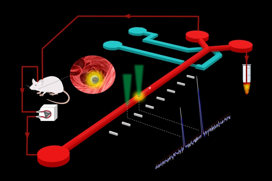
The device draws blood from a mouse, routes it through a microfluidic chip, and looks for tumor cells that glow under laser light. In the mouse model used by the researchers, tumor cells carry a fluorescent marker, so the system can spot them while they are mixed into the enormous background of normal blood cells.
When the chip detects a glowing CTC, it diverts a tiny volume of blood - about 100 nanoliters - into a collection tube. The rest of the blood is returned to the same awake mouse, which is the key design feature that makes serial sampling possible.
Why microfluidics matters here
A good analogy is a highway toll lane that can pull one specific car out of heavy traffic without stopping the whole road. Microfluidics does something similar with blood: it guides very small volumes through precisely engineered channels so the system can identify and isolate rare cells with minimal disruption.
That precision matters because CTCs are vanishingly rare. Finding them is like trying to catch a few specific grains of sand in a moving river, and standard methods often require much larger blood draws or end the experiment after a single collection point.
What researchers can learn from repeat sampling
The main advance is not just that the team can capture CTCs, but that they can do it over time from the same animal. Instead of comparing different mice at different disease stages, researchers can track how tumor cells appear, change, and possibly become more dangerous as cancer progresses.
After collection, the sample can be purified further to separate individual CTCs from the thousands of other blood cells that came along in the tiny diverted fraction. That opens the door to studying single tumor cells one by one, which is important because not all CTCs are alike; some may be harmless passengers, while others may be the ones most capable of starting metastases.
A better window into metastasis
Metastasis is the process by which cancer spreads from its original site to distant organs. Scientists know CTCs are part of that process, but the timing and biology of their transition from primary tumor cells to metastatic founders has been difficult to observe directly.
By enabling collection over days and weeks, the MIT system gives researchers a way to examine that transition as it happens. That could help answer practical questions such as when CTC numbers rise, whether their traits shift during treatment or progression, and which cells are most likely to survive the trip through the bloodstream.
What makes this different from older approaches
Previous approaches often forced a tradeoff between cell access and animal survival. Researchers could analyze blood or tumor tissue at a single moment, but they usually could not keep returning to the same mouse to build a time series of CTC behavior.
This system changes that by acting more like a closed loop than a one-way harvest. Blood leaves the mouse, passes the detection and sorting step, and then goes back, allowing longer observation periods and making it easier to connect CTC biology to the evolving disease in one individual animal.
Why This Matters
Cancer researchers often need better tools before they can get better answers, and this is a clear example. If metastasis is the deadliest part of cancer, then technologies that let scientists inspect the cells responsible for that spread - repeatedly, with timing and context - could sharpen both basic research and future drug development.
The work does not by itself produce a new treatment, and the source describes it in a mouse model, not in patients. But methods like this can reveal which tumor cells matter most, how they change over time, and when intervention might be most effective, which are exactly the kinds of insights that eventually shape diagnostics and therapies.
What comes next
The broader promise is a more dynamic view of cancer, one that follows movement and change instead of relying on static snapshots. As researchers use tools like this to connect microfluidic engineering with the biology of metastasis, they may be able to build a clearer map of how cancer spreads - and where that process can be interrupted.