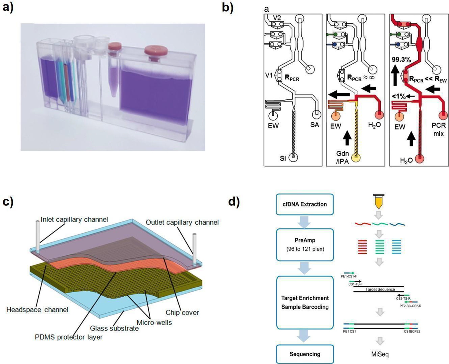
Integrated lab-on-a-chip microfluidic systems are being developed to do a difficult but clinically important job: find tiny traces of cancer in ordinary body fluids such as blood, saliva, and urine. The review behind this article surveys how these miniature devices can isolate, enrich, and analyze cancer biomarkers—measurable signs of disease such as circulating tumor cells, extracellular vesicles, nucleic acids, and proteins. The appeal is easy to grasp. Instead of relying only on a tissue biopsy, which can be invasive and hard to repeat, a liquid biopsy aims to monitor cancer through a simple sample that can be collected again and again. But the science is tough because many of these biomarkers are extraordinarily rare, especially early in disease. The authors argue that combining multiple sample-preparation and detection steps on one chip could help solve that problem by reducing sample loss, speeding analysis, and making tests more practical outside specialized labs. Their overall message is measured rather than breathless: the field has produced promising prototype systems, yet major technical and clinical hurdles still stand between elegant devices and routine medical use. Even so, the review suggests that steady engineering progress and closer collaboration across disciplines could make liquid biopsy a more reliable tool for early cancer detection and treatment monitoring.
Why rare biomarkers are so hard to catch
Cancer sheds a mix of material into the bloodstream and other fluids. Among the best-known examples are circulating tumor cells, or CTCs, which are whole cancer cells that break away from a tumor; extracellular vesicles, or EVs, which are tiny membrane-wrapped packages released by cells; and pieces of DNA, RNA, or proteins linked to tumor activity.
The challenge is scale. The review notes that CTCs can be present at concentrations roughly seven orders of magnitude lower than white blood cells in blood, especially in early cancer. In everyday terms, that is like trying to find a few specific grains of sand on a crowded beach before you can even start asking what those grains mean.
What a lab-on-a-chip actually does
A microfluidic device moves and manipulates very small volumes of liquid through channels smaller than the width of a human hair. Think of it as a miniaturized plumbing system built onto a chip, where pumps, filters, mixers, and sensors are shrunk down and connected so that one small sample can be processed step by step.
In cancer diagnostics, the goal is not just to detect a molecule but to run an entire workflow on one platform. That can include separating target biomarkers from everything else in the sample, concentrating them so they are easier to measure, and then analyzing them with optical, electrical, or biochemical readouts. Integration matters because every transfer between tubes or instruments risks losing some of the very material researchers are trying to detect.
Two main strategies: biochemical and biophysical capture
The review groups isolation and enrichment methods into two broad families: biochemical and biophysical. Biochemical approaches use affinity, meaning they rely on molecules that specifically bind to a target—much like a lock and key. Antibodies are a common example, because they can be designed to latch onto proteins found on the surface of tumor cells or vesicles.
Biophysical methods, by contrast, sort particles based on traits such as size, shape, deformability, density, or electrical properties. A simple analogy is a kitchen sieve that separates large pieces from small ones, though the real devices are much more precise. These methods can be useful when a biomarker does not carry a clean molecular label or when researchers want to avoid bias toward only one biological marker.
Why integration is the key engineering idea
What makes this review notable is its focus on integrated systems rather than stand-alone chips that perform only one task. A useful liquid-biopsy device has to do several things well in sequence: accept a real-world sample, handle contaminating cells and proteins, enrich the rare signal, and then produce an analysis that clinicians can trust. Building all of that into one compact format is the central engineering challenge.
Integration offers obvious advantages if it works. It can reduce hands-on steps, shorten turnaround time, lower reagent use, and make results more reproducible by standardizing the workflow. For clinics, a single automated chip is far more attractive than a patchwork of manual procedures that require specialized training and expensive lab infrastructure.
Promising prototypes, but not a solved problem
The review’s conclusion is cautiously optimistic. The authors say the promising performance of existing detection systems shows that clinically meaningful liquid biopsies are attainable, which is an important distinction: the field has proof-of-concept success, but not yet universal clinical readiness.
Several obstacles still slow progress. Biomarkers differ widely from one cancer type to another, and even from one patient to another, so a system tuned for one target may miss another. Devices must also cope with messy, variable samples from actual patients, not just idealized laboratory specimens, while maintaining sensitivity, specificity, and consistent manufacturing quality.
From research tool to clinical test
Moving from an impressive prototype to a routine diagnostic test usually depends on more than clever chip design. A clinical assay needs validation in large patient groups, clear evidence that the result changes medical decisions, and workflows that fit into hospitals and diagnostic labs. It also has to be robust enough to produce the same answer across operators, sites, and batches of devices.
This is where the review emphasizes multidisciplinary collaboration. Engineers can design fluidic architectures, chemists can optimize capture surfaces, biologists can clarify which biomarkers matter most, and clinicians can define what level of performance is actually useful in patient care. Without that chain of cooperation, elegant technology may remain stuck at the demonstration stage.
Why This Matters
If integrated microfluidic liquid-biopsy systems mature, they could change how cancer is detected and monitored. Repeated tissue biopsies are often difficult, uncomfortable, or impossible, while fluid sampling is generally simpler and less invasive. That opens the door to tracking disease progression, treatment response, and possibly early signs of relapse more frequently.
The broader significance is access. A well-designed chip-based platform could shrink sophisticated cancer testing into a format that uses smaller samples, less equipment, and more automation, which may help extend advanced diagnostics beyond top-tier research centers. The review does not claim that this future has already arrived, but it makes a strong case that integrated systems are one of the most practical routes toward it.
What comes next
The field now appears to be in a transitional phase: past the point of simple concept papers, but still working through the hard details that determine clinical adoption. Future progress will likely depend on devices that can handle multiple biomarker types, maintain accuracy with real patient samples, and fit within regulated diagnostic workflows. If those pieces come together, lab-on-a-chip systems could turn liquid biopsy from a promising idea into a routine part of cancer care.