Researchers have outlined a more systematic way to design self-assembling immunotherapies by linking the chemistry of peptide antigens to how immune signals physically come together. The work focuses on therapeutic complexes built from positively charged peptide antigens and negatively charged nucleic acid-based modulatory cues, two ingredients that naturally attract each other like opposite poles of a magnet. By adding molecular dynamics simulations—computer models that track how atoms move and interact over time—the team examined how small design changes alter the assembly process at a very fine scale. They found that peptides with more positive charge, and especially those anchored with the amino acid arginine, formed more electrostatic contacts than comparable peptides using lysine or carrying less charge. The study also showed that total charge was not the whole story: where that charge sits along the peptide changed binding strength as well. Experimental tests backed up the simulations, with surface plasmon resonance measurements and primary immune cell studies showing that these structural choices shaped how strongly the components bound and how cells responded. The broader message is practical: if researchers can predict how immune signals assemble before they build them, they may be able to design immunotherapies more rationally for autoimmune disease and other targets. That kind of molecular-level map could make nanomaterial-based immune treatments less trial-and-error and more precise.
Using self-assembly to tune immune signals
The project starts from a basic challenge in immunotherapy: the immune system does not respond only to what signal it sees, but also to how that signal is packaged and presented. The authors describe a platform in which peptide antigens are modified with cationic residues, meaning positively charged amino acids, and then combined with anionic nucleic acid-based cues, which carry negative charge.
That pairing lets the components self-assemble, meaning they organize themselves into complexes without external construction step by step. A simple analogy is snapping together building blocks that are shaped to fit and also pull on each other electrically. In this case, the “fit” comes from the peptide design, while the “pull” comes from charge.
Why simulations mattered here
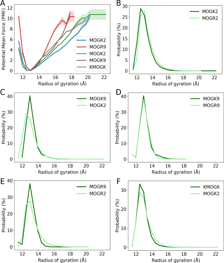
The team used temperature replica exchange molecular dynamics, a simulation method that helps researchers sample many possible molecular arrangements rather than getting stuck in just one. Think of it as watching the same molecules explore multiple versions of the same dance floor under slightly different conditions, then comparing the moves they settle into most often.
That approach let the researchers count specific interactions during self-assembly, including hydrogen bonds and salt bridges. Hydrogen bonds are weak attractions that help stabilize biomolecules, while salt bridges are stronger electrostatic interactions between opposite charges. Both matter because they can influence whether a therapeutic complex forms tightly, loosely, or not at all.
What changed when peptide design changed
Across a library of peptide sequences associated with antigens mistakenly targeted in autoimmune disease, the simulations revealed clear patterns. Peptides carrying higher cationic charge formed more electrostatic interactions during self-assembly than peptides with lower positive charge.
The identity of the anchored amino acid also mattered. Peptides anchored with arginine formed more electrostatic interactions than those anchored with lysine, even though both amino acids are positively charged. That distinction is important because it shows that not all positive charges behave the same way once molecules begin to pack together.
Charge distribution was as important as charge amount
The study did not stop at total charge. The researchers also found that the distribution of charge across the peptide influenced binding affinity, or how strongly the self-assembled immune cues stuck together.
They tested this with surface plasmon resonance, a technique that measures molecular binding in real time by detecting tiny changes at a sensor surface. In everyday terms, it works a bit like measuring how firmly two pieces of Velcro catch as they touch and pull apart. Those experiments showed that where the charges were placed along the peptide changed the strength of the interaction, alongside the choice of arginine or lysine.
Cell studies connected structure to immune signaling
The computational and biophysical findings were then checked in in vitro primary cell studies, meaning experiments performed with living cells outside the body. These tests used the same antigen designs examined in the simulations and binding experiments.
The cell results tracked with the earlier observations. Immune signaling was sensitive to total charge, charge distribution, and the identity of the anchored amino acid residues within the therapeutic complexes. In other words, the same molecular features that changed assembly and binding also changed how cells interpreted the signal.
What this says about autoimmune antigens
The peptide library in this study represented sequences that are mistakenly attacked during autoimmune disease. That gives the work a concrete therapeutic context: these are not abstract model materials, but antigens relevant to disorders in which the immune system turns against the body’s own tissues.
By comparing several related peptide designs rather than just one example, the team could ask a more useful question for therapy development: which molecular edits consistently improve assembly and signaling behavior? The answer appears to depend on a combination of factors, not a single design rule.
Why This Matters
Many immunotherapies are still optimized through repeated testing, where researchers make a material, measure its effects, adjust it, and try again. This study points toward a more predictive approach by tying microscopic interactions—hydrogen bonds, salt bridges, and charge placement—to measurable outcomes such as binding affinity and cell signaling.
That matters because self-assembled immune materials can be difficult to control if their behavior is treated as a black box. A better understanding of nanomaterial-immune interactions could help scientists adapt the same platform to different peptide antigens and disease targets without starting from scratch each time. For autoimmune disease in particular, precision is crucial: the goal is to retrain or redirect immune responses, not simply stimulate them more strongly.
From trial-and-error toward rational design
The central contribution of the work is not a single finished therapy, but a design logic for building them. By combining simulations, binding measurements, and cell-based readouts, the researchers created a framework for asking how a peptide’s sequence shapes the assembled complex and how that complex shapes immune behavior.
That kind of framework could become increasingly useful as immunotherapy developers work with broader sets of antigens and more complex biomaterials. If researchers can forecast how specific molecular features will influence assembly before moving into the lab, they may be able to build safer, more targeted immune therapies with fewer rounds of guesswork.