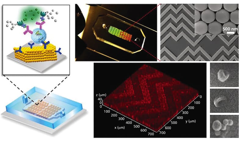
A University of Kansas team has developed a lab-on-a-chip designed to detect signs of cancer from a tiny droplet of blood plasma, the liquid component of blood. The device focuses on exosomes, extremely small particles released by cells that can carry molecular clues about their cell of origin. Its central advance is a three-dimensional nanoporous structure patterned like a herringbone, a familiar branching shape seen in nature. That structure helps move exosomes toward the chip's sensing surface, where capture probes can recognize them. In testing with clinical samples from ovarian cancer patients, the chip detected the presence of cancer using only a minuscule amount of plasma. The researchers frame ovarian cancer as a model for a platform that could potentially be adapted to other cancers and neurodegenerative diseases. If the design proves reliable in broader testing, it could support less invasive, lower-cost forms of testing often called liquid biopsies. The work illustrates how carefully shaping fluid flow at microscopic scales can make a diagnostic sensor more effective.
A Blood Test Built Around Tiny Cell Messages
A liquid biopsy is a test that looks for disease-related material in blood or another body fluid rather than requiring a tissue sample collected through surgery or a needle procedure. In this case, the target is the exosome, a nanoscale membrane-bound particle that cells release into their surroundings.
Exosomes can contain biological material associated with the cells that produced them. That makes them attractive diagnostic targets, but their tiny size and limited abundance in a small sample create a practical challenge: a sensor must bring enough of them into contact with its detection surface to register a useful signal.
Why Contact With the Sensor Is Hard
The University of Kansas device addresses a basic physical bottleneck known as mass transfer. In plain terms, mass transfer is the movement of particles through a fluid to a place where they can be measured, much like trying to get every grain of tea to reach a strainer rather than simply drift around a cup.
For a chip sensor, exosomes must travel through liquid and approach a surface carrying probes, molecules designed to recognize and capture a specific target. As particles get very close to that surface, a thin layer of liquid remains between them, and the resulting hydrodynamic resistance makes it harder for the particles to close the final distance.
A Herringbone Structure at the Nanoscale
The chip's key feature is a three-dimensional, nanoporous herringbone pattern that both mixes biological material and improves sensing. Nanoporous means it contains extremely small openings, while the herringbone arrangement refers to repeated angled branches resembling the bones of a fish.
According to the description of the device, this structure drains liquid from the narrow gap between a particle and the sensing surface. That action pushes exosomes into hard contact with the surface, increasing the chance that the probes there can recognize and capture them.
Testing With Ovarian Cancer Samples
The researchers tested the microfluidic chip using clinical samples from ovarian cancer patients. A microfluidic device controls very small volumes of liquid through miniature channels, effectively putting a laboratory workflow onto a compact chip.
The reported result was detection of cancer in a very small quantity of plasma. The source does not provide the number of samples, the diagnostic accuracy, or a comparison with established clinical tests, so the result should be understood as a demonstration of the chip design rather than evidence that it is ready to replace existing diagnostic practice.
What Could Make This Format Useful
The design could matter because it tries to solve a diagnostic problem without requiring a large blood draw or a complex, large-scale instrument. A chip that can work with small plasma volumes may be especially useful where sample availability, repeated monitoring, or testing cost are important practical constraints.
The source also states that these microfluidic chips could be cheaper and easier to manufacture than comparable designs. Manufacturing simplicity does not automatically guarantee low clinical cost, since validation, sample preparation, readout equipment, and regulatory requirements all matter, but a simpler device architecture can be an important starting advantage.
Beyond One Cancer Type
Ovarian cancer served as the model system for proving the chip's design, not as the only intended use. The same approach could potentially be applied to breast and colorectal cancers, as well as neurodegenerative diseases, if researchers can identify the relevant exosome-associated markers and build suitable capture probes.
That distinction is important. The herringbone structure is a way of improving the physical delivery of particles to a sensor, while the probes on the sensor determine what biological signal the device is actually looking for. A platform may therefore be adaptable, but each new disease application still requires careful development and validation.
Why This Matters
Diagnostic tests often succeed or fail on an unglamorous detail: whether the target molecules or particles can reliably reach the part of the device meant to detect them. This chip treats fluid mechanics as a core part of the diagnostic design, using nanoscale geometry to overcome the resistance created by a thin liquid gap near the sensing surface.
For patients, the appeal of a liquid biopsy is straightforward: a blood-based test could be less invasive than obtaining tissue directly. For clinicians and researchers, a device that concentrates and captures exosomes efficiently could create another route to studying disease signals that circulate outside the cells themselves.
What Comes Next
The next step is likely to be broader clinical evaluation that measures how consistently the chip performs across more samples and against standard diagnostic methods. If that testing supports the early result, the University of Kansas design could help make exosome-based analysis more practical, showing how a small patterned surface can turn a droplet of plasma into a potentially useful source of diagnostic information.