Fluorescence imaging is one of the most useful ways to watch biology happen inside microfluidic devices, the tiny chip-based systems that move and process liquids through channels thinner than a millimeter. But there has long been a frustrating tradeoff: researchers can either image a large area of a chip or zoom in with enough light sensitivity to see faint fluorescent signals, yet rarely both at once. A study on a tandem-lens macroscope tackles that problem by describing an imaging setup built to capture a wide field of view while preserving the brightness needed for fluorescence work. The approach is aimed at experiments where scientists need to monitor many channels, chambers, or droplets across a single device without stitching together multiple images. That matters because microfluidic platforms are increasingly used for cell studies, diagnostics, and chemical analysis, where events unfold across an entire chip rather than at one tiny spot. By adapting an optical design more often associated with low-magnification, high-light-collection imaging, the work offers a practical route to making whole-device fluorescence readouts easier. In effect, the study shows how researchers can better match their microscope to the scale of the chip they are studying. For labs building or using biochips, that could make experiments faster, more quantitative, and easier to interpret.
A better match for chip-scale imaging
Traditional fluorescence microscopes are excellent for cellular detail, but they are not always ideal for microfluidics. Many chips are physically large compared with the narrow viewing window of standard microscope objectives, forcing users to scan across the device or accept only partial information.
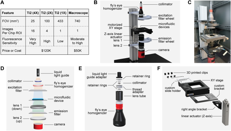
The tandem-lens macroscope is designed for the opposite priority. Instead of maximizing magnification, it emphasizes a large field of view and efficient light collection, which are exactly what researchers need when fluorescent signals are spread across many regions of a chip.
What a tandem-lens macroscope does
A macroscope is an optical system for imaging relatively large objects at low magnification. In a tandem-lens arrangement, two camera lenses are placed face to face so that one lens collects light from the sample and the other relays it to the detector, creating a bright image over a wide area.
This matters for fluorescence because fluorescent labels are often dim. The more light the system can gather, the easier it is to detect labeled cells, molecules, or droplets without requiring excessively long exposure times or harsh illumination that could damage samples.
Why fluorescence in microfluidics is challenging
Microfluidic devices often pack a lot of function into a compact footprint: multiple channels, reaction chambers, traps, or mixing regions can all exist on one chip. Researchers may want to compare activity across the whole device at once, such as whether a reagent reaches every channel evenly or whether cells respond differently in different zones.
With a narrow-field microscope, that kind of overview is cumbersome. Scientists may need to move the stage, capture many frames, and stitch them together later, which adds time and can introduce errors if the sample changes during acquisition.
The practical advantage of seeing more at once
The value of a wide-field fluorescence setup is not just convenience. Capturing the entire device in one image can improve experimental consistency because every region is recorded under the same illumination and at the same moment in time.
That is especially important in dynamic experiments. In droplet microfluidics, for example, droplets move quickly; in cell assays, fluorescent responses can rise and fall over minutes; and in chemical gradients, concentrations can shift continuously across a chip.
What the study contributes
The reported system shows that a tandem-lens macroscope can be adapted for large-area fluorescence imaging of microfluidic devices. Rather than treating chip imaging as a scaled-down version of conventional microscopy, the work recognizes that biochips have their own optical requirements and benefits from an imaging geometry built around them.
That design choice can lower the barrier for experiments that need whole-chip observation. It also opens the door to more quantitative measurements, since researchers can compare signals across the device without worrying that different regions were captured at different times or under slightly different focus conditions.
Where this could be useful
Applications are broad. Labs working on cell sorting, single-cell analysis, organ-on-a-chip systems, or high-throughput screening could use this kind of imaging to monitor many assay sites simultaneously.
It could also help in diagnostic chip development, where engineers need to verify that signals appear in the correct chambers and with the expected intensity. A large-field fluorescence view is well suited to troubleshooting device design, validating prototypes, and checking whether fluid handling is working as intended.
Why This Matters
Biochip technology often advances through a combination of better device design and better ways to read those devices out. This study focuses on the second part of that equation, showing that imaging hardware can be just as important as the chip itself in determining what researchers are able to measure.
More broadly, the work reflects a shift toward tools tailored to the realities of microfluidics rather than borrowed unchanged from traditional microscopy. As chip-based experiments become larger, more multiplexed, and more automated, imaging systems that can see an entire device clearly and sensitively will become increasingly valuable.
Looking ahead
The tandem-lens macroscope will not replace high-magnification microscopy when researchers need fine structural detail. But for many chip-level fluorescence assays, it offers a compelling compromise between coverage and sensitivity that standard setups struggle to provide.
The next step is likely integration with faster cameras, automated analysis, and custom illumination schemes that make full-device imaging even more powerful. If that happens, large-field fluorescence could become a standard way to monitor microfluidic experiments in real time, helping turn complex lab-on-a-chip systems into more reliable research and diagnostic tools.