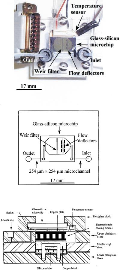
Researchers developed a glass-silicon microchip module that can pull white blood cells out of a tiny drop of whole blood and then run a polymerase chain reaction, or PCR, on the captured cells without moving the sample to a separate instrument. In the reported setup, the device handled less than 3 microliters of human blood, isolated the cells using a built-in filter, and then amplified a 226-base-pair region of the human coagulation Factor V gene. That matters because sample preparation is often the slow, messy part of genetic testing: blood has many components, and the DNA-containing white blood cells need to be separated from the far more numerous red blood cells before analysis. By combining those steps on one module, the team showed a path toward smaller, simpler testing systems that use very little sample. The work also highlights why microchips became so attractive for biology in the first place: they can be etched with fine structures that guide fluids and cells almost like roads and checkpoints on a map. Here, those structures included a series of 3.5-micrometer weir-type filters, small barriers designed to trap larger nucleated cells while letting other material pass through. The result was a proof of principle that blood preparation and DNA amplification do not have to be separate workflows. Instead, they can be linked inside one compact platform, which is exactly the kind of integration needed for faster point-of-care and low-volume genetic tests.
How the chip works
The core idea is easy to picture. Imagine pouring mixed pebbles and sand through a carefully sized grate: the larger pieces get caught, while the smaller ones keep moving. In this microchip, the "pebbles" are white blood cells, which contain nuclei and therefore genomic DNA, and the filtration structures are etched into silicon at feature sizes of about 3.5 micrometers.
The filter itself was described as a silicon dam spanning the flow chamber, creating a series of weir-type barriers. As whole blood moved through the channel, these barriers retained the white blood cells needed for downstream analysis. Red blood cells, which are much more abundant in blood and lack nuclei, were not the target for the DNA test.
From blood drop to PCR
After priming the microchip with phosphate-buffered saline, or PBS, containing a small amount of Triton X-100 to remove air and reduce handling problems, the researchers introduced less than 3 microliters of human whole blood. The sample came from a fasting volunteer and was reported to contain about 5,400 white blood cells per microliter and 4.8 million red blood cells per microliter.
That huge imbalance is exactly why blood is a challenging starting material. The useful DNA for this assay sits inside a relatively small population of cells, buried in a fluid dominated by red blood cells. The chip's job was to enrich the useful fraction directly inside the device, rather than requiring a separate benchtop purification step.
Why integration is the key advance
Once the blood had been loaded, the team pumped 100 microliters of PCR assay mixture through the chip at 9.85 microliters per minute to complete the isolation process and set up amplification. In plain terms, the chip did not just catch cells; it also became the place where those cells were processed for DNA testing. That is a meaningful shift from conventional workflows, where samples are usually transferred between tubes, filters, and thermal cyclers.
PCR, short for polymerase chain reaction, is a method that copies a chosen DNA sequence over and over until there is enough material to detect. You can think of it like photocopying one page from a huge library until that page becomes easy to study. In this case, the chosen page was a 226-base-pair segment of the human Factor V gene, and the DNA template came directly from white blood cells isolated on the filter section of the chip.
Built on earlier microfilter designs
The work did not appear in isolation. The authors positioned the module within a line of research exploring different microchip geometries for handling human cells, including arrays of posts, tortuous channels, comb-shaped filters, and earlier weir-type filters. Different silicon microfilters ranging from 3 to 10 micrometers had also been fabricated across microchannels and evaluated for filtration efficiency.
That design history matters because it shows this module was not just about proving PCR could happen on a chip. It was also about refining the physical architecture needed to make blood sample preparation reliable at very small scales. Glass and silicon were especially useful materials because they could be patterned precisely, allowing the team to tailor channel shapes and filter dimensions to the behavior of cells in flowing blood.
What the module demonstrates
The main demonstration was direct amplification from DNA released out of white blood cells captured inside the device. In other words, the same microchip that performed the separation step also supported the genetic assay. For a lab workflow, removing a transfer step is more important than it sounds, because every transfer adds time, contamination risk, and sample loss.
The tiny sample volume is another striking feature. Working with less than 3 microliters of whole blood suggests the approach could be useful when sample is scarce, such as finger-prick testing, neonatal screening, or repeated monitoring where minimizing blood draw matters. The study did not claim to solve every challenge of integrated diagnostics, but it showed that essential front-end and amplification steps can live in one module.
Why This Matters
For nonexperts, the significance is simple: most DNA tests are not limited by the chemistry of copying DNA, but by the practical hassle of getting a clean, usable sample into the reaction. Blood is full of cells and molecules that can complicate analysis, so a device that sorts the right cells and runs PCR in one place could make testing smaller, faster, and easier to automate. That is especially relevant to biochip development, where the long-term goal is to shrink complex lab procedures into portable systems.
This approach also points toward broader integrated assays. The authors noted that the module should be adaptable to additional assay types, which means the same basic platform could eventually support other genetic targets or upstream sample-handling tasks. In the bigger picture, the study helped define a practical roadmap for lab-on-a-chip systems: combine physical cell handling with molecular detection, and do both with as little sample and user intervention as possible.
What comes next
The next steps for technologies like this are usually about robustness, not just proof of concept. A useful clinical device would need to handle more patient-to-patient variability, operate reproducibly outside a specialist lab, and support targets beyond a single gene fragment. Even so, this microchip module captured the central promise of biochips early and clearly: if you can isolate the right cells and amplify their DNA on the same tiny platform, you are much closer to turning a complicated lab procedure into a practical test.