Source: Biomarker Research, by Zhiting Liu; Yingyu Zhou; Jia Lu; Ting Gong; Elena Ibáñez; Alejandro Cifuentes; Weihong Lu. AI-generated summary by biochip.com, published . Not independently reviewed.
Microfluidic biosensors are becoming one of the most promising tools for spotting cancer earlier, when treatment has the best chance of working. The core idea is simple: instead of testing a large tube of blood or other body fluid all at once, these devices move tiny amounts of liquid through miniature channels, giving sensors tighter control over what they measure. In the source article, researchers describe how this approach can improve the detection of biomarkers, the molecular signals linked to disease, including small extracellular vesicles, or sEVs, that circulate in body fluids. One example focuses on early-stage breast cancer, where a microfluidic system uses magnetic beads and antibodies to isolate vesicles carrying Glypican-1, also called GPC-1. By combining careful fluid handling with biochemical capture steps, the platform aims to detect rare targets that might be missed by conventional methods. The broader argument is that microfluidics does more than miniaturize a lab test: it can boost sensitivity, shorten response times, and improve specificity by increasing contact between the target molecule and the sensor. The article also highlights how microfluidics can be paired with electrochemical, fluorescent, and nanoparticle-based detection methods, creating a flexible toolkit for cancer diagnostics. Taken together, the story is about a shift toward smaller, smarter tests that may one day make early cancer screening faster, less invasive, and more precise.
How the microfluidic approach works
A useful way to picture a microfluidic device is to think of a highly organized road system for droplets and cells. Instead of letting everything mix in a large container, the device guides tiny fluid volumes through narrow channels, where each step can be controlled with much more precision.
That control matters because cancer biomarkers in blood, plasma, or other body fluids can be present at extremely low levels. If the system can steer those targets directly toward a sensing surface, it increases the odds of capturing and measuring them before they get lost in background noise.
Using magnetic beads to catch rare vesicles
The source article describes a biosensor built around magnetic beads modified with a CD63 antibody. An antibody is a protein that recognizes a specific molecular feature, and CD63 is commonly used as a marker on the surface of small extracellular vesicles, tiny membrane-bound particles released by cells.
In the breast cancer example, the goal is to capture sEVs carrying GPC-1 from patient plasma. Plasma is the liquid part of blood, and because it can contain many different particles, the challenge is to isolate the right vesicles cleanly enough to support an accurate test.
Why droplet microfluidics helps
The platform uses droplet microfluidics, which means the sample is split into tiny droplets that act like miniature reaction chambers. This helps separate and organize interactions so that, as the article explains, each target sEV can be captured by a single magnetic bead after mixing and magnetic separation.
That one-bead, one-target logic is important because it reduces confusion during measurement. If too many particles clump together, the signal can become harder to interpret, but isolating individual complexes makes the readout cleaner and potentially more reliable.
Building a readable signal
After the vesicle-bead complexes form, they are recognized again by detection antibodies carrying enzymatic markers. This is similar to adding a second lock-and-key step: the first antibody grabs the target, and the second helps label it so the device can generate a measurable signal.
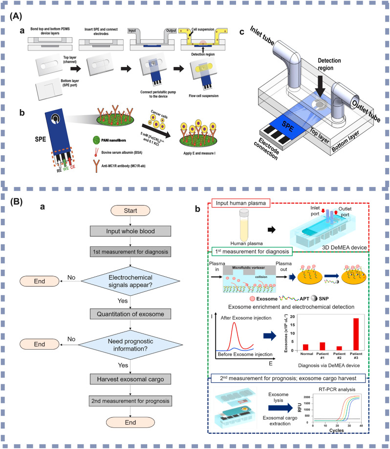
Those labeled complexes are then introduced into the microfluidic device for detection. The source notes that multiple electrodes can be arranged inside the microfluidic channel as an electrode array, turning the chip into a more complete sensing system rather than a simple capture platform.
Better sensitivity, speed, and specificity
According to the article, bringing microfluidics into these biosensors has markedly improved several core performance measures, including detection limit, response time, and specificity. Detection limit refers to the smallest amount of a biomarker that can still be reliably measured, which is crucial for early cancer because the signal may be very faint.
The improvement comes from precise control of fluid dynamics, meaning how liquids move and mix at very small scales. When a device can manage that movement carefully, it increases contact between the sensor and the analyte, the substance being measured, while also limiting unwanted interactions that create false signals.
Different sensing strategies on the same kind of chip
The article emphasizes that microfluidics is not tied to one detection method. Instead, it acts like a versatile chassis that can be combined with several sensing technologies, each with different strengths for cancer biomarker analysis.
Electrochemical sensors, for instance, are highlighted for their high sensitivity. In practical terms, they translate chemical interactions into electrical signals, making them especially useful when biomarker concentrations are very low and every bit of signal matters.
Fluorescence-based detection is another major option. Here, biomarkers are tagged with molecules that emit light, allowing highly specific detection and making these microfluidic systems suitable for precise diagnostics even when the target concentration is extremely small.
The article also points to the role of nanoparticles, which can expand what these devices can do. Because nanoparticles have unusual optical, electrical, and surface properties, they can amplify signals or improve target capture, helping biosensors detect cancer-associated molecules with greater efficiency.
Why This Matters
Early cancer diagnosis often depends on finding subtle molecular clues before a tumor causes obvious symptoms. A microfluidic biosensor that can detect rare vesicles or other biomarkers in accessible body fluids could support less invasive testing, potentially using blood or similar samples rather than tissue collected through more difficult procedures.
That matters not just for patient comfort, but also for timing. If clinicians can identify disease earlier and monitor it more easily, they may be able to make treatment decisions sooner, follow response over time, and catch recurrence with more precision.
What comes next
The source article presents microfluidic biosensors as a key direction for cancer diagnostics because they combine miniaturization, selective capture, and sensitive readout in a single platform. The remaining challenge is turning these promising designs into robust tools that work consistently across real clinical samples, where body fluids contain a messy mix of cells, proteins, and particles.
Still, the trajectory is clear. As microfluidics is integrated more tightly with electrochemical readouts, fluorescent labels, and nanoparticle-assisted sensing, these devices may move closer to routine screening and monitoring, bringing early detection into settings far beyond the traditional laboratory.