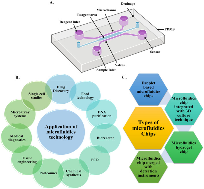
Microfluidic chips are tiny lab systems etched with channels so small they can guide droplets, cells, and chemicals with extreme precision, and a recent review argues that these devices are moving from clever engineering tools toward core platforms for biomedical research and medicine. The big idea is simple: shrink parts of a laboratory onto a chip and you can run experiments faster, with less sample and less reagent, while controlling conditions more tightly than in many conventional setups. The review surveys how these chips are designed, what materials and layouts matter most, and where the field is heading in diagnostics, therapy, and disease modeling. It highlights a broad shift from basic fluid handling toward systems that can mimic organs such as the lung, kidney, skin, brain, and gut. It also points to droplet microfluidics—methods that package tiny volumes of fluid like miniature test tubes—as a powerful route for targeted drug delivery and high-throughput screening. In one example discussed by the authors, researchers used a droplet-based microfluidic approach to build pH-sensitive and magnetic drug carriers aimed at colorectal cancer treatment. Taken together, the article presents microfluidic chips not as one device, but as a flexible toolkit for making biology easier to measure, model, and manipulate.
How a Microfluidic Chip Works
You can think of a microfluidic chip as a city map for liquids. Its tiny microchannels connect in carefully planned ways, with inlets and outlets linking the chip to pumps, samples, and detectors outside the device.
Once fluid enters the chip, those channels can split, merge, mix, or hold it in place for reactions. Because the volumes are so small, heat and chemicals move quickly, which often speeds up experiments and reduces the amount of material needed.
Why the Design Matters
The review emphasizes that chip performance depends heavily on design strategy. Channel geometry, the arrangement of inputs and outputs, and the interface between the larger outside world and the tiny on-chip environment all shape how fluids behave.
That matters because fluid behavior changes at small scales. Effects that are easy to ignore in a beaker—such as surface forces and the exact shape of a channel—can dominate on a chip, so good design is less about miniaturizing a standard lab protocol and more about rethinking it for a microscopic setting.
What Makes These Devices Attractive
The appeal of microfluidic chips comes from a practical combination of speed, efficiency, and parallelization. They typically use less reagent, consume smaller samples, and can perform several operations at once, which is especially valuable when working with expensive chemicals or scarce patient material.
Their small size can also make reactions faster. Shorter distances inside the chip mean molecules do not have to travel as far, so mixing and physicochemical responses can happen on a timescale that is hard to match in larger systems.
From General Platforms to Organ-on-Chip Systems
One of the review's central themes is the rise of organ-on-chip technology, which aims to recreate key features of human organs inside a microfluidic device. A simple analogy is a flight simulator: it does not reproduce an entire airplane or the whole sky, but it reproduces the conditions that matter most for testing performance and behavior.
In the same way, an organ-on-chip tries to capture important aspects of tissue physiology, such as fluid flow, mechanical forces, and cell-to-cell interactions. The review points to devices modeled on the lung, kidney, skin, brain, and gut, all of which are being used to study biology in environments that are more realistic than flat cell cultures in a dish.
Why Organ Mimics Are Useful
These systems matter because many diseases and treatments depend on physical context, not just on the identity of the cells involved. Lung tissue stretches as we breathe, kidney tissue experiences filtration-like flow, and gut tissue interacts with nutrients and microbes, so a chip that reproduces those conditions can reveal effects that standard cell culture may miss.
That makes organ-on-chip platforms attractive for both diagnostics and therapeutics. In the biomedical setting described by the review, they are being used to probe disease mechanisms, test drug responses, and build models that could eventually narrow the gap between preclinical experiments and what happens in patients.
Droplet Microfluidics and Targeted Delivery
The review also highlights droplet microfluidics, a branch of the field that creates tiny, discrete droplets inside another fluid. The everyday analogy is a conveyor belt of perfectly uniform bubbles, except each bubble can act like its own miniature reaction vessel.
That high level of control is useful for making drug carriers and screening many conditions quickly. Because each droplet can be formed with nearly identical size and composition, researchers can build particles or capsules with more predictable properties than they might get from bulk mixing.
A Colorectal Cancer Example
As one case study, the review cites work by Maher and colleagues on colorectal cancer treatment using droplet-based microfluidics. The researchers designed multifunctional transporters that were both magnetic and pH-responsive, meaning they were built to react to acidity changes that can differ between healthy tissue and tumor environments.
According to the review, the platform used pH-sensitive magnetic microspherical carriers loaded with the anticancer agents 5-fluorouracil and curcumin. Those drugs were chosen because their combination had shown a synergistic effect, and the cargo was stacked on permeable silicon nanoparticles and magnetic bacterial iron oxide nanowires to create a more sophisticated delivery system.
Why This Matters
The broad significance of this review is that it frames microfluidic chips as more than miniaturized plumbing. They are becoming decision-making tools for biomedicine: ways to model disease, sort out how drugs behave, and build targeted delivery strategies that are difficult to assemble with conventional methods.
That shift could help researchers move faster while wasting less material, an important practical advantage in both academic labs and clinical development. If a chip can mimic critical features of an organ or produce highly controlled therapeutic carriers, it can reduce trial-and-error and make experiments more informative from the start.
What Comes Next
The future perspective in the review is one of expansion rather than replacement. Microfluidic chips are unlikely to eliminate every traditional lab method, but they are steadily becoming better at handling complex biological questions that need precision, realism, and scale at the same time.
The next phase will likely depend on improving robustness, standardization, and real-world integration with diagnostics and therapy workflows. If those pieces come together, microfluidic chips could move from being specialized research tools to routine parts of how scientists study organs, design treatments, and test medical ideas before they reach patients.