Source: Biosensors, by Khaled Alsabbagh; Tim Hornung; Achim Voigt; Sahba Sadir; Taleieh Rajabi; Kerstin Länge. AI-generated summary by biochip.com, published . Not independently reviewed.
Researchers have described a microfluidic impedance biosensor chip that can detect proteins without adding fluorescent or chemical labels, using cardiac troponin I as a test case. The device combines a standard glass microscope slide, gold electrodes, and a small PDMS channel, a rubbery polymer commonly used to guide tiny amounts of liquid in lab-on-a-chip systems. What makes the design notable is the sensing layer on the gold surface: the team compared conventional thiol-based coatings made from small hydrocarbons with coatings built from thiolated DNA, short DNA strands that self-assemble into an organized film. In their tests, the DNA-based layer did a better job of blocking unwanted protein sticking while still allowing the target protein to be recognized by an antibody. That matters because false signals from random protein adsorption are one of the biggest obstacles in biosensors meant for real biological samples. When the researchers flowed in human serum albumin, a common blood protein that often causes background interference, the chip showed almost no impedance change at 1000 ng/mL. But when they applied troponin I at 1 ng/mL, the electrical readout shifted clearly, suggesting the device could pick up clinically relevant biomarker levels within minutes.
How the chip is built
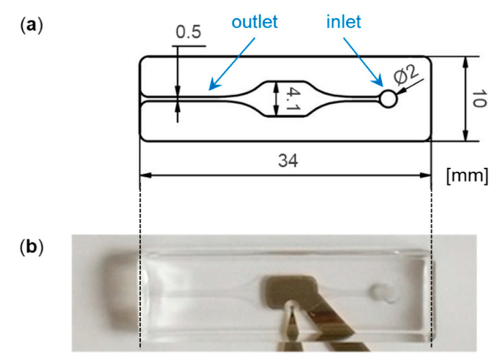
The sensor is built on a simple physical stack. A glass slide serves as the base, gold electrodes are sputtered onto it, and a polydimethylsiloxane microchannel sits on top to direct fluid across the sensing area.
That architecture is familiar in microfluidics because it is practical and relatively low cost. Glass provides a stable surface, gold is easy to modify chemically, and PDMS is easy to mold into narrow channels that use only tiny sample volumes.
What impedance sensing actually measures
The chip reads out changes through impedance, which is a measure of how strongly a system resists electrical current when an alternating signal is applied. A useful everyday analogy is traffic on a road: if more obstacles appear, cars move differently, and you can infer what is blocking the path without seeing each object directly.
On this sensor, protein binding changes the electrical behavior at the electrode surface. Instead of tagging the protein with a dye or enzyme, the researchers watch how the surface itself alters the current, creating a label-free measurement.
Why the surface chemistry matters so much
For a biosensor like this, the sensing layer is the gatekeeper. It has to hold the antibody in place, allow enough charge transfer for a measurable signal, and stop unrelated proteins from sticking and creating false positives.
The team tested self-assembled monolayers, or SAMs, on the gold electrodes. These are tightly packed molecular coatings that form spontaneously when the right sulfur-containing molecules, called thiols, bind to gold, a bit like bristles arranging themselves neatly on a brush head.
DNA-based monolayers outperformed hydrocarbon coatings
The study compared SAMs made from thiolated aromatic hydrocarbons with SAMs made from thiolated oligonucleotides, short DNA fragments modified so they can anchor to gold. The DNA-based coatings showed better overall performance in the final sensor setup.
Specifically, they were better at suppressing nonspecific adsorption of human serum albumin or HSA, one of the most abundant proteins in blood. At the same time, they still supported specific binding of troponin I through the matching antibody attached to the surface.
How the target protein was captured
To make the chip selective, the researchers attached the corresponding antibody to the SAM using carbodiimide chemistry. In plain terms, this is a common coupling method that acts like a molecular connector, linking chemical groups on the surface to chemical groups on the antibody.
That matters because the antibody provides the biological recognition step. Troponin I is not detected just because it is present in solution; it is detected because the antibody is designed to bind that specific cardiac protein much more strongly than unrelated proteins.
What the tests showed
The key comparison in the report is straightforward. A sample containing HSA at 1000 ng/mL produced negligible impedance changes, while a troponin I sample at 1 ng/mL caused a significant shift in the Nyquist plot, a standard way to visualize impedance data.
That split is important because it suggests the signal is driven by specific recognition rather than random fouling. In a practical setting, a biosensor must ignore abundant background proteins while responding clearly to a lower-concentration biomarker of interest.
Why troponin I is a meaningful test case
Troponin I is one of the most specific serum biomarkers used to help diagnose myocardial infarction, better known as a heart attack. When heart muscle is damaged, troponin leaks into the bloodstream, and clinicians use its presence and concentration as a key piece of diagnostic evidence.
That makes it a demanding and clinically relevant target for a chip-based assay. A sensor that can identify troponin quickly, with small sample volumes and minimal preparation, could fit well into point-of-care testing where speed matters.
Why This Matters
The most interesting part of this work is not just that the chip detected a protein, but that it did so with a surface designed to solve a classic biosensor problem: nonspecific binding. In real samples such as blood or serum, stray proteins can coat a sensor like dust on a camera lens, blurring the signal and making a test unreliable.
By showing that DNA-based SAMs can reduce that background while preserving electrical responsiveness and antibody capture, the study points to a practical route for more robust label-free sensors. The reported measurement time of only a few minutes also hints at applications beyond the lab bench, where fast turnaround is often as important as analytical sensitivity.
What comes next
The results are presented as promising rather than final, and that is the right way to read them. A strong next step would be testing the chip in more complex clinical samples and against a wider panel of interfering proteins to see how well the surface chemistry holds up outside controlled conditions.
Even so, the work offers a clear design lesson for biochip developers: the smartest part of a sensor is often the invisible molecular layer on top. If that layer can combine selective capture, low fouling, and clean electrical readout, compact chips like this one may become more useful for rapid protein testing in medicine.