Engineers in Prof. Amin Arababian’s laboratory have built a microfluidic system designed to find rare cells in blood at very high speed without attaching fluorescent or chemical labels to them first. The idea targets a stubborn problem in liquid biopsy, the practice of looking for disease-related cells or signals in a blood sample instead of taking tissue directly from a tumor or other organ. In the team’s design, blood is funneled through many tiny channels on a lab-on-a-chip device, and cells are identified by how they interact with specially prepared regions inside those channels. That matters because standard cell-sorting methods often require extra preparation steps, added reagents, and bulky hardware, all of which can slow testing and raise costs. The new approach aims to replace some of that complexity with a label-free readout based on flow behavior, electrical sensing, and software that can tell one channel from another. According to the source, the system is meant for ultra-high-throughput analysis, meaning it could inspect very large numbers of cells quickly while still tracking signals at the single-cell level. The proposed uses range from circulating tumor cell detection in liquid biopsies to fetal cell analysis and whole-blood cell counting. If the platform performs as intended in practice, it could help move sophisticated cell analysis closer to point-of-care settings, where testing happens near the patient rather than in a large centralized lab.
How the chip is supposed to work
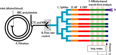
A good way to picture the device is as a highway system shrunk onto a chip. One blood sample enters, then gets divided again and again into many smaller lanes so the system can process many cells in parallel instead of forcing everything through a single bottleneck.
The source describes a first separation step in which smaller red blood cells, or RBCs, and platelets are split away from larger white blood cells, or WBCs, and circulating tumor cells, or CTCs. That size-based filtering is useful because the cells researchers usually want to inspect in a liquid biopsy are much rarer and often hidden within a huge background of ordinary blood components.
Detection without labels
The key technical claim is label-free detection. In plain terms, that means the system tries to recognize cells directly, without first tagging them with fluorescent dyes, magnetic beads, or antibody-linked markers that traditional sorting systems often rely on.
Instead, each channel contains “interaction zones,” which are functionalized segments of the microfluidic pathway. These regions are designed so that a cell’s particular set of surface biomarkers changes how easily it moves through the channel, effectively turning cell identity into a measurable change in flow behavior.
That is a bit like placing different kinds of Velcro patches along a narrow hallway and then watching which shoes slow down at which spot. Scientifically, the “Velcro” is the surface chemistry in each interaction zone, and the slowdown reflects how strongly a cell’s outer molecules interact with that chemistry.
Why throughput is such a hard problem
Finding rare cells in blood is not just a biology challenge; it is also an engineering challenge. If a device uses hundreds of microfluidic channels at once, the electronics needed to read every lane separately can quickly become too large for the chip and too messy to interpret cleanly.
The source says this is one of the main obstacles the new system tries to solve. Large numbers of parallel channels can create interference and a wiring problem, where the support electronics begin to overwhelm the compact advantages of a lab-on-a-chip platform.
Shared electronics and channel “ID numbers”
To get around that bottleneck, the team developed an electronics sharing scheme paired with signal-processing software. Rather than dedicating a full set of hardware to every single channel, the system assigns each channel an effective “ID number” that software can decode.
That means the chip can identify where a target cell was detected while using only a minimal number of electronic channels. In practical terms, it is similar to hearing many instruments through a smaller number of microphones and then using software to figure out which sound came from where.
This matters because miniaturized biology tools often fail not in the fluidics, but in the supporting hardware needed to read them out. If the decoding method works reliably, it could make massively parallel microfluidic analysis more realistic outside a specialized engineering lab.
Electrical sensing inside the channel
The device does not rely only on interaction zones. The source also describes electrodes placed between those zones to identify cells of interest through impedance measurements and transit-time analysis.
Impedance is a way of measuring how much a material resists an electrical signal. In a microfluidic channel, different cells disturb that signal in different ways, and the time a cell takes to pass between sensing points can add another clue about its size, shape, or interactions with the channel surface.
Combining these readouts could make the platform more informative than a simple yes-or-no detector. It suggests the system is trying to build a richer signature for each cell, using both biochemical interactions and electrical behavior to improve specificity.
Potential uses beyond cancer
Liquid biopsy is the most obvious application because clinicians and researchers want better ways to isolate rare circulating tumor cells from blood. Those cells can potentially support downstream testing, such as genetic analysis, without the need for an invasive tissue procedure every time a patient is monitored.
But the same platform could also be useful for fetal cell detection and routine blood analysis. Because the chip first sorts cells by size and then examines specific surface-marker interactions, it could be adapted for different cell types depending on how the channels are functionalized.
The source also notes that after detection, targeted cells can be sorted and collected for downstream analysis, including fluorescence-activated cell sorting, or FACS, and DNA sequencing. That is important because counting a suspicious cell is only the first step; clinicians and researchers often want to study that cell in much greater detail afterward.
Why This Matters
Many of today’s best cell-analysis systems are powerful but expensive, reagent-heavy, and tied to centralized laboratories. A microfluidic device that can perform specific, multiplexed, single-cell analysis without the usual labeling step could lower cost and simplify workflows at the same time.
There is also a speed advantage in principle. When a system can process many channels in parallel and reduce sample-prep steps, it becomes easier to imagine faster turnaround for tests that depend on finding a tiny number of meaningful cells among millions of ordinary ones.
Just as important, the design points toward point-of-care use, meaning testing closer to where patients are seen. That does not guarantee immediate clinical adoption, but it highlights the broader goal: shrinking advanced cell analysis into a compact platform that is easier to deploy.
What to watch next
The source presents the architecture and intended advantages, but the next questions are practical ones: how accurately the chip detects target cells, how well it handles real-world blood samples, and how easily the system can be manufactured and integrated into clinical workflows. Those details will determine whether the platform remains an elegant engineering concept or grows into a useful diagnostic tool.
Even so, the project captures a larger shift in bioengineering. Researchers are trying to combine microfluidics, electrical sensing, and software into compact systems that do more with less hardware, less sample preparation, and less time—an approach that could reshape how rare-cell detection is performed in both research and medicine.