AI-generated summary by biochip.com, published . Not independently reviewed. Source: Nanotheranostics, by Qiao-ru Guo; Ling-ling Zhang; Ji-fang Liu; Zhen Li; Jia-jun Li; Wen-min Zhou; Hui Wang; Jing-quan Li; Da-yu Liu; Xi-yong Yu; Jian-ye Zhang.
Microfluidic chips are being developed as compact tools that could help doctors both find cancer earlier and test treatments more realistically. Instead of relying only on tissue biopsies or imaging, these devices can analyze tiny amounts of blood or other samples for cancer biomarkers—measurable signs of disease such as circulating tumor cells, circulating tumor DNA, exosomes, non-coding RNA, proteins, and metabolites. The source article describes how one platform can bring several of these jobs onto a single chip, using microscopic channels to sort cells, detect molecules, and even model how tumors respond to drugs. That matters because conventional screening can be invasive, expensive, and difficult to repeat often. A chip-based approach promises something closer to a lab shrunk onto a glass slide: faster handling of samples, tighter control of fluids, and the possibility of running several tests at once. The review also points to specific research reports in journals including Lab on a Chip, Scientific Reports, Biosensors and Bioelectronics, and Analytical Chemistry that show how these systems are being used for cell capture, biomarker detection, and drug screening. Taken together, the work suggests microfluidics is moving from a niche engineering tool toward a more practical cancer platform that links diagnosis with treatment decisions.
What the chip is trying to do
A microfluidic chip moves very small volumes of liquid through tiny channels, a bit like routing traffic through a dense network of miniature roads. Because researchers can precisely control where cells and molecules go, the chip can be built to isolate rare targets, mix reagents, and measure signals in a single device.
In cancer, that is especially useful because the most informative material is often scarce. A blood sample may contain only a small number of circulating tumor cells, or fragments of tumor DNA mixed into a huge background of normal material, so the technology has to be selective as well as sensitive.
The biomarkers researchers are chasing
The source highlights several major classes of cancer biomarkers. These include circulating tumor cells, or CTCs, which are whole cancer cells that break away from a tumor and enter the bloodstream; circulating tumor DNA, or ctDNA, which consists of genetic fragments shed by tumors; and exosomes, tiny membrane-bound particles released by cells that carry molecular cargo.
It also points to non-coding RNA, proteins, and metabolites. Non-coding RNA is RNA that does not make proteins but can still regulate cell behavior, while metabolites are small molecules produced during normal and abnormal cell activity. Detecting these markers accurately can help with early diagnosis and may also help grade cancers, meaning estimate how aggressive a tumor is likely to be.
Why microfluidics appeals to cancer diagnostics
Standard cancer workups often depend on tissue biopsy or medical imaging. Those tools remain essential, but biopsies are invasive and imaging may miss subtle molecular changes that appear before a tumor is obvious on a scan.
Microfluidic systems aim to fill that gap by working with small, repeatable liquid biopsies—tests based on blood or other body fluids. The appeal is practical: less sample, less manual handling, and the ability to combine separation and detection steps that would otherwise require several instruments and trained staff.
On-chip detection of circulating tumor cells
One major application is on-chip CTC detection. Think of it like panning for gold in a river, except the gold flakes are rare cancer cells moving among millions of blood cells. The chip can use channel geometry, flow behavior, or surface chemistry to help isolate those rare cells.
The source cites a 2017 Lab on a Chip study describing a microchip device for the separation and dual detection of prostate cancer cells and protein biomarkers. That combination is important because it moves beyond a simple yes-or-no cell count and toward a richer readout that links cell capture with molecular information from the same sample.
Measuring how cancer cells behave
Microfluidic chips are not only collection devices; they can also probe cell behavior. The source references a 2015 Scientific Reports paper on microfluidic cytometric analysis of cancer cell transportability and invasiveness, meaning how easily cells move and how capable they are of penetrating surrounding tissue.
An everyday analogy is a stress test for cars on different roads. By forcing cells through controlled microchannels, researchers can see how deformable and mobile they are, traits that may relate to metastatic potential—the ability of cancer to spread to other parts of the body.
Using chips to test drugs in a realistic tumor setting
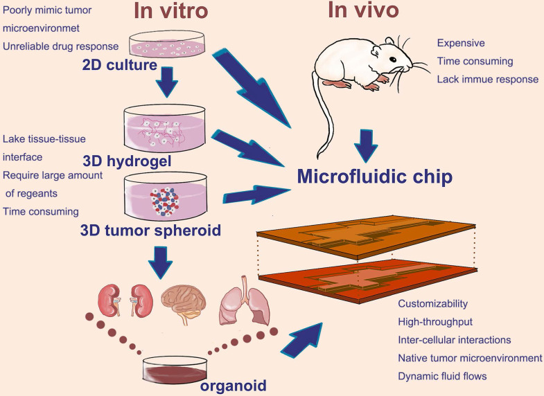
Another thread in the source is treatment testing. A 2017 paper in Biosensors and Bioelectronics described a microfluidic platform for drug screening in a 3D cancer microenvironment, which tries to mimic the physical and chemical surroundings of a tumor more closely than flat cells in a dish.
That matters because cancer cells often behave differently in three dimensions than they do on a standard lab plate. A microfluidic device can expose them to gradients of nutrients, oxygen, and drugs, offering a more realistic look at whether a therapy works before it reaches a patient.
Toward personalized treatment monitoring
The source also cites a 2019 Analytical Chemistry paper titled Personalized Drug Efficacy Monitoring Chip. The title alone points to a larger ambition in the field: not just diagnosing cancer, but testing which therapy is most likely to help a specific patient.
This is where the "multifunctional" idea becomes especially important. A single chip that can detect biomarkers, evaluate tumor-like cells, and monitor drug response could shorten the path between finding a cancer signal and making a treatment decision tailored to that signal.
Why This Matters
The biggest promise of these chips is integration. In today’s cancer care, diagnosis, molecular profiling, and therapy selection are often separated into different workflows, sometimes using different samples collected at different times. Microfluidic platforms suggest a more connected model, where one small sample can generate several layers of information.
That does not mean the technology is ready to replace standard care across the board. But the review makes clear that researchers are steadily building devices that can capture rare cells, detect molecular markers, and model treatment response with increasing sophistication. If those tools become robust enough for clinics, they could make cancer monitoring less invasive and more frequent.
What comes next
The next challenge is translation: turning elegant lab prototypes into dependable clinical tools. For that to happen, researchers will need to show that multifunctional chips work consistently across real patient samples, not just controlled experiments, and that the results improve decisions doctors already make. Still, the direction is clear. By compressing many lab functions into a small, precisely engineered device, microfluidic chips are giving cancer diagnosis and treatment a shared platform rather than two separate tracks.