A team at the Georgia Institute of Technology has developed a microchip designed to catch one of cancer’s most dangerous travelers: circulating tumor cell clusters, or groups of cancer cells that break away from a tumor and move through the bloodstream. These clusters are thought to play an outsized role in metastasis, the process by which cancer spreads to other organs and becomes much harder to treat. The new device, called the Cluster-Well, aims to isolate those clusters quickly and without relying on chemical labels that can alter or miss important cells. Instead, it combines the fine control of microfluidics, which manipulates tiny amounts of fluid through microscopic channels, with a filtration strategy that traps clusters in tiny wells. Researchers say that design could make it easier not only to detect these cells from a routine blood sample, but also to preserve them for later study. That matters because even a single circulating tumor cell can reveal clues about a patient’s disease, from how aggressive the cancer is to which treatments might work best. If the approach holds up in broader testing, it could help doctors monitor metastatic disease earlier and more precisely than is possible with standard imaging alone.
Why circulating tumor cells matter
Most cancer deaths are linked not to the original tumor, but to metastasis. That process begins when tumor cells detach, enter the bloodstream, and eventually seed new tumors elsewhere in the body.
Scientists have studied circulating tumor cells, or CTCs, for years as a potential liquid biopsy marker. A liquid biopsy is a test that looks for cancer-related material in blood rather than requiring a tissue sample from surgery or a needle biopsy.
Why clusters are especially important
Single tumor cells in the blood are useful, but clusters may be even more revealing. The Georgia Tech researchers note that a lone cell often struggles to survive the harsh conditions of circulation, while clusters are much more resilient.
That durability may make clusters especially effective at establishing metastases. In other words, finding and analyzing clustered cells could offer a sharper view of when a cancer is becoming more dangerous and how it may be spreading.
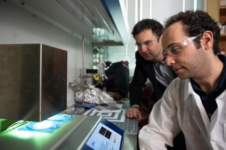
How the Cluster-Well works
The new chip is designed to capture these clusters using meshed microwells, tiny well-like structures with built-in filtering features. As blood moves through the device, clusters are trapped in those wells while remaining accessible for imaging and follow-up analysis.
This design is notable because it blends two strengths that are often difficult to combine. Microfluidic chips are highly precise and can position cells in controlled ways, while membrane filtration is efficient at separating particles by size; the Cluster-Well tries to do both at once.
A label-free approach
Another key feature is that the method is label-free. Many cell-isolation techniques depend on molecular tags that bind to specific proteins on the cell surface, but those methods can miss cells that do not express the expected markers.
By avoiding labels, the chip may capture a broader and less biased sample of tumor cells and clusters. That is important in cancer because tumor cells are notoriously variable, even within the same patient, and the most dangerous cells may not always fit a standard molecular profile.
What the researchers found
According to the source material, the team observed hundreds of circulating tumor cells in clusters in blood samples from ovarian cancer patients, with some of those cells still alive. That detail is especially striking because viable cells can be studied much more deeply than damaged or dead ones.
Living CTCs could potentially be examined for drug sensitivity, genetic changes, and other traits tied to treatment response. Preserving the biology of these cells, rather than simply counting them, could make the test more useful for guiding real clinical decisions.
From blood draw to treatment insight
The promise of this approach is straightforward: a simple blood sample could reveal how a patient’s cancer is behaving in near real time. Doctors currently rely heavily on scans and occasional tissue biopsies, both of which can miss rapid changes or be difficult to repeat frequently.
A chip-based assay could provide a less invasive way to watch metastatic disease evolve. If clinicians can see that dangerous clusters are increasing, changing, or carrying particular markers, they may be able to adjust therapy sooner.
Why This Matters
This work points toward a future in which metastatic cancer is monitored more like a chronic condition, with repeated sampling rather than isolated snapshots. That would be a meaningful shift because metastasis can progress quickly, and treatment decisions are often made with incomplete information.
The ability to isolate intact cell clusters also opens a window into basic cancer biology. Researchers still do not fully understand why some clusters survive, why others fail, or which ones are most likely to colonize new organs, and tools like this could help answer those questions.
What comes next
The research was presented by Utkan Demirci Sarioglu’s group in a study titled High Throughput, Label-free Isolation of Circulating Tumor Cell Clusters in Meshed Microwells, published in Nature Communications. As with many promising biomedical devices, the next steps will likely include broader validation across more patients and cancer types, along with efforts to show that the chip improves care in a real clinical setting.
Still, the concept is compelling: instead of waiting for metastasis to become obvious on a scan, clinicians may someday be able to detect and study the cells driving it through a blood test. If that happens, tools like the Cluster-Well could help make cancer treatment faster, more targeted, and better matched to the biology of each patient’s disease.