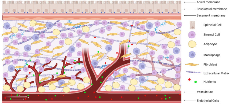
Organ-on-chip devices are emerging as a practical way to test whether new chemicals are likely to harm people before those compounds ever reach the clinic or the market. In a recent perspective on new approach methodologies, or NAMs, researchers argue that these miniature systems could fill a major gap left by standard lab models such as flat cell layers and animal tests, both of which often predict human toxicity poorly. The core idea is simple: instead of asking isolated cells or whole animals to stand in for a human organ, build a tiny device that recreates key parts of the organ’s structure and function. That can mean arranging cells in separate compartments, adding an extracellular matrix—the protein-rich scaffold cells normally sit in—and flowing nutrients through the device the way blood would in the body. The authors say the science behind these systems has advanced quickly, but adoption in ordinary tissue culture labs has not kept pace. In their view, the main bottlenecks are not a lack of biological realism, but weak regulatory standards and poor fit with existing lab workflows. Their perspective lays out what organ-on-chip systems need to become more predictive, more reproducible, and easier for analytical chemists to use in real toxicity testing.
Why current toxicity models fall short
Toxicity testing still leans heavily on two familiar tools: cells grown as thin monolayers in dishes and animal models. Both are useful, but both simplify biology in ways that can hide or distort how a human organ will respond to a new chemical entity, the term used for a newly developed compound.
A flat layer of cells is a bit like judging how a city works by looking only at one street from above. You may see some activity, but not the traffic flow, the neighborhoods, or the infrastructure connecting them. In the same way, monolayer cultures miss the three-dimensional organization, cell-to-cell separation, and dynamic fluid movement that shape real tissue behavior.
What makes an organ-on-chip different
Organ-on-chip devices aim to recreate small but meaningful pieces of organ function inside a controlled lab platform. These systems are usually microengineered devices that hold living cells in carefully designed spaces, allowing researchers to mimic how tissues are organized and how they experience mechanical and chemical signals.
The perspective highlights several features the authors see as essential. The first is anatomically relevant structure: cells should be arranged in ways that reflect actual tissue architecture rather than simply spread onto plastic. That includes cellular compartmentalization, where different cell types are kept in distinct but connected regions, and the use of extracellular matrices that give cells the cues they need to behave more naturally.
The importance of flow, time, and sampling
Another key requirement is continuous perfusion, meaning a steady flow of fluid through the device. Think of it as replacing a stagnant pond with a moving stream. In living organs, cells constantly receive fresh nutrients and chemical signals while waste products are carried away, so a perfused chip can support longer and more realistic studies.
This matters especially for toxicity testing because harmful effects do not always appear immediately. Some chemicals injure tissues only after prolonged exposure, or after cells alter the chemical over time. A device that can run for longer periods under flowing conditions gives researchers a better chance of seeing these delayed or cumulative effects.
The authors also stress a more practical point that often gets less attention: chips must generate enough sample material for quantitative analysis. Analytical chemists do not just want to see whether cells look stressed under a microscope. They often need measurable outputs—metabolites, proteins, signaling molecules, or other chemical readouts—in amounts large enough to analyze reliably and at scale.
Why analytical chemists care
This is where the perspective becomes especially relevant for analytical chemistry. Organ-on-chip systems are not only biological models; they are also measurement platforms. Their value depends on whether researchers can collect reproducible data from them and connect those data to meaningful toxicological outcomes.
The authors argue that device design should support routine sampling and compatibility with standard analytical methods. That sounds mundane, but it is crucial. A beautifully realistic chip is far less useful if it cannot easily plug into the instruments, sample preparation steps, and workflows already used in tissue culture and toxicity labs.
Progress in fabrication and accessibility
One reason the field is gaining traction is that fabrication and analysis methods are becoming more accessible. Earlier generations of organ-on-chip devices could feel specialized and difficult to build, limiting them to engineering-heavy labs with niche expertise.
The perspective points to recent advances that are making these devices more tractable for mainstream tissue culture laboratories. In other words, the technology is moving closer to a point where labs can adopt it without rebuilding their entire operation. That shift may be just as important as any single biological improvement, because a model only helps if researchers can actually use it consistently.
The real barriers to adoption
The authors make a striking argument: organ-on-chip adoption is limited less by biology than by infrastructure. They contend that the devices already offer strong biological relevance, but they still face two systemic problems—few standardized regulatory benchmarks and limited compatibility with established experimental workflows.
Regulatory benchmarks matter because companies and testing labs need clear rules for what counts as valid evidence. If one chip design uses different materials, cell layouts, sampling schemes, and endpoints from another, it becomes hard to compare results across laboratories or convince regulators that the data should inform safety decisions. Without standards, even promising devices can remain stuck in pilot studies.
Workflow compatibility is the second hurdle. Most toxicity labs are built around familiar protocols for monolayer culture and animal studies. If a chip platform requires unusual handling, custom analysis pipelines, or difficult interpretation, researchers may hesitate to switch even when the biology is better.
Why This Matters
The bigger significance of organ-on-chip technology is not simply that it looks more sophisticated than older models. It is that better models could lead to better early decisions about which chemicals are safe enough to advance and which should be stopped sooner. That could save time, reduce cost, and lower reliance on animal testing while improving the chances that toxicity signals seen in the lab will match what happens in humans.
NAMs combine in vitro methods, which test biology outside the body, with in silico methods, which use computation to model outcomes. Organ-on-chip devices fit into that broader shift toward testing strategies that are more human-relevant and data-rich. If the field can standardize performance criteria and make these systems fit naturally into existing analytical workflows, chips may become less of a specialist tool and more of a routine part of safety science.
For now, the message from the perspective is clear: the promise of organ-on-chip devices does not hinge only on making them more lifelike. It also depends on making them measurable, reproducible, and usable in ordinary labs. The next phase of progress will likely come from that intersection of engineering, cell biology, and analytical chemistry, where a better model is only as good as the evidence it can consistently produce.