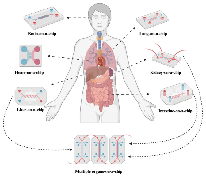
Organ-on-a-chip technology is moving from a clever lab concept toward a practical tool for drug development, toxicity testing, and disease research. These devices, also called microphysiological systems, use tiny channels to grow living human cells under controlled conditions that resemble the body more closely than a standard petri dish can. The core idea is simple: instead of testing a drug in flat cell cultures that miss key features of real tissue, researchers build a miniature environment where cells experience fluid flow, mechanical forces, and neighboring cell types similar to those in an organ. The review article highlights how this approach is being applied across health science, especially in studies of drug response, safety, and disease modeling. It also points to the rise of linked systems, such as gut chips connected with liver or kidney chips, to study what happens after a drug is absorbed and then processed by the body. A major example is the gut-on-a-chip, where researchers have recreated motions like intestinal squeezing and structures like villi, the tiny finger-like folds that help the intestine absorb nutrients. Together, these advances suggest that organ chips could help researchers predict human biology earlier and more accurately, while reducing some of the limits of animal models and conventional cell culture.
How organ chips work
An organ-on-a-chip is a small microfluidic device, meaning it uses tiny channels to move fluids in a highly controlled way. You can think of it like a miniature plumbing system for cells: instead of water moving through pipes, nutrients, drugs, and signaling molecules flow past living tissues.
That flow matters because cells behave differently when they are exposed to motion, pressure, and changing chemical conditions. In the body, cells are never sitting still on a flat plastic surface, so organ chips try to recreate the physical cues that tell cells how to grow, specialize, and interact.
Why researchers are using them for drug development
Drug development often fails because early test systems do not reflect human biology well enough. A compound may look safe in a simple cell culture but behave very differently once it is absorbed, metabolized, or exposed to a more realistic tissue barrier.
The review describes organ chips as a promising answer to that problem. By maintaining cells under more life-like conditions, these platforms can help researchers study how drugs move through tissues, how strongly they affect target cells, and whether they damage organs that were not meant to be hit.
Better ways to study toxicity
Cellular toxicity assessment means asking a basic but critical question: does a treatment harm healthy cells while trying to help a patient? Traditional models can miss subtle injury signals because they do not reproduce the architecture and stress patterns of real tissues.
Organ chips improve that picture by letting scientists observe cells in a dynamic environment over time. That is especially useful for toxicity studies, where timing, dose, and repeated exposure can all shape whether a compound is tolerated or dangerous.
The gut-on-a-chip as a leading example
Among the many organ chip designs, the gut-on-a-chip stands out because the intestine is both mechanically active and central to how the body handles food, microbes, and many medicines. If a normal cell dish is like looking at a city from a single rooftop, a gut chip is closer to watching traffic move through streets, bridges, and checkpoints.
The review traces this progress back about a decade to early systems designed to mimic peristalsis, the wave-like squeezing motion that pushes material through the intestines. In 2012, researchers reported a gut-on-a-chip that reproduced this physical motion, giving intestinal cells a more realistic mechanical environment than static culture can provide.
A year later, researchers showed that applying shear stress and cyclic strain could drive cultured human intestinal cells to form villi. Shear stress is the frictional force created by flowing fluid, and cyclic strain is repeated stretching and relaxation; together, these cues helped create the finger-like structures that support absorption and barrier function in the real intestine.
Why villi and barrier function matter
The formation of villi is not just a visual milestone. These structures increase surface area and help the intestine decide which molecules can pass into the body and which should stay out, making them central to both nutrition and drug absorption.
That barrier behavior is one reason gut chips are so valuable for pharmaceutical research. If a drug cannot cross the intestinal lining properly, or if it damages that lining on the way through, researchers need to know early, before moving deeper into development.
Connecting organs to follow a drug's path
One of the more important advances discussed in the review is the move from single-organ chips to linked systems. In these setups, a gut chip is paired with chips representing organs such as the liver or kidneys, which allows researchers to study what happens after a drug passes through the intestine.
This matters because the body handles medicines as a chain of events, not isolated steps. A drug absorbed through the gut may be changed by the liver into a more active compound, an inactive one, or even a toxic byproduct, and kidney function may then shape how long that substance remains in circulation.
By connecting these systems, researchers can examine drug metabolism and organ safety in sequence rather than as separate experiments. That gives a more realistic picture of how an orally delivered treatment might behave in a person.
Disease modeling and personalized medicine
The review also emphasizes disease modeling, where the goal is not just to test a compound but to recreate aspects of illness in a controlled device. That can help researchers study how a disease starts, how it changes tissue behavior, and which interventions might interrupt it.
This approach is especially attractive in areas where standard models oversimplify complex biology. Because organ chips can combine physical forces, tissue barriers, and multiple cell types, they may capture disease features that are hard to reproduce in conventional cultures.
The same logic extends to personalized medicine, which aims to tailor care to an individual patient. In principle, chips built with patient-derived cells could help reveal why one person responds well to a treatment while another experiences side effects or little benefit.
Why This Matters
Organ-on-a-chip systems matter because they try to close one of the biggest gaps in biomedical research: the distance between a simple lab model and a living human body. They are not full replacements for animal studies or clinical trials, but they offer a more human-relevant testing ground at a stage where better predictions can save time, money, and failed effort.
That is particularly important for drug safety. If researchers can spot absorption problems, off-target toxicity, or harmful metabolic effects earlier, they may avoid pushing weak candidates forward and focus resources on compounds with a better chance of helping patients.
The growing focus on gut-related biology also makes these tools timely. As researchers pay more attention to gut axes, meaning the intestine's connections with other organs and body-wide disease processes, realistic gut models become more useful for understanding conditions that extend far beyond digestion.
What comes next is likely to be more integration rather than less: more linked organs, more disease-specific models, and more attempts to make chip behavior match the body even more closely. If that trend continues, organ-on-a-chip platforms could become a standard bridge between early discovery and real-world medicine, helping researchers ask better questions before treatments ever reach a patient.