AI-generated summary by biochip.com, published . Not independently reviewed. Source: Pigment Cell & Melanoma Research, by Xiangjie Jin; Yuhan Zhang; Yuantai Zhu; Zhiyuan Zhang; Chaoji Shi.
Mucosal melanoma, a rare form of melanoma that arises in the body’s moist internal linings rather than the skin, remains one of the hardest melanomas to study and treat. Unlike the more familiar cutaneous melanoma, which is linked to sun exposure, mucosal melanoma has different genetic features, often appears later, and tends to respond poorly to standard therapies. The review article summarized here argues that one of the field’s biggest problems is not just the biology of the disease, but the lack of good models that let researchers test ideas quickly and realistically. Scientists have built a growing toolkit of laboratory systems, including cell lines, patient-derived xenografts, organoids, and animal models, but each captures only part of the disease. That matters because mucosal melanoma is both rare and highly diverse, making it difficult to design one-size-fits-all treatment strategies. The authors also point to a newer class of tools: virtual models that use computation, artificial intelligence, and multi-omics data to simulate how tumors, immune cells, and drugs interact. Their central message is simple: progress will likely come not from one perfect model, but from combining physical and digital systems to close the gap between lab findings and patient care.
A cancer with different rules
Mucosal melanoma develops in tissues such as the nasal passages, mouth, gastrointestinal tract, and female genital tract. Because these sites are hidden and symptoms can be vague, the disease is often found only after it has already advanced.
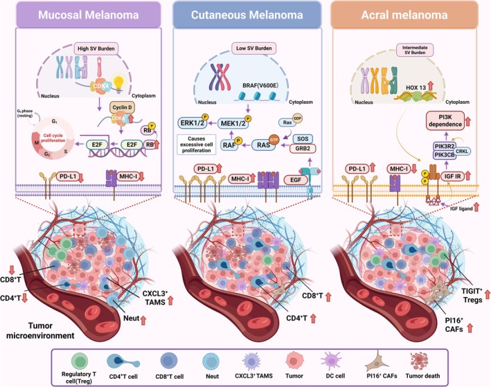
The review notes that this cancer is especially important in Asian populations, where it represents a larger share of melanoma cases than it does in Western populations. It also behaves differently from skin melanoma at the molecular level, which helps explain why treatments borrowed from cutaneous melanoma often work less well here.
Why current treatment falls short
Doctors treating mucosal melanoma face a frustrating reality: there is still no widely accepted standard regimen tailored specifically to this disease. Surgery, radiation, targeted therapy, and immunotherapy are all used, but outcomes remain poor for many patients.
Part of the problem is that mucosal melanoma appears to have a distinct and often immunosuppressive microenvironment—the local ecosystem of immune cells, structural cells, and signaling molecules around the tumor. A useful analogy is bad soil around a plant: even if you use the right seed, growth will not follow the usual pattern because the environment changes everything. In cancer, that altered environment can blunt the effect of therapies that rely on the immune system to attack tumor cells.
What preclinical models can teach researchers
To understand a difficult cancer, scientists need stand-ins that behave enough like the real tumor to test drugs and mechanisms. Traditional cell lines, which are cancer cells grown in dishes over long periods, are still useful because they are relatively easy to handle and can support repeatable experiments.
But cell lines have clear limits. Over time, they can drift away from the original tumor, and they usually miss the complexity of the surrounding tissue, immune system, and three-dimensional structure that shape how a cancer grows and resists treatment.
Models built from patient tumors
More advanced systems try to preserve more of the original disease. Patient-derived xenografts, or PDXs, are made by implanting a patient’s tumor into an animal, usually a mouse, so researchers can observe growth and treatment response in a living body.
Patient-derived organoids, often called PDOs, are another promising option. These are miniature three-dimensional tumor structures grown from patient tissue; you can think of them as tiny architectural mock-ups that preserve more of the building’s layout than a flat blueprint does. Because organoids can retain important features of the source tumor, they may help researchers compare drug responses across different patients more realistically than standard cell cultures can.
The problem of rarity
Even the best model systems run into a basic obstacle: there simply are not many mucosal melanoma samples to work with. The cancer is rare, and the tumors themselves are heterogeneous, meaning that one patient’s disease may differ substantially from another’s in location, genetics, and immune behavior.
That scarcity limits the number of available cell lines, PDXs, and organoids, and it makes standardization difficult. If every lab studies a small and slightly different set of models, it becomes harder to compare results or build a clear picture of which findings are broadly true and which apply only to a narrow subset of cases.
Why animal comparisons still matter
The review also highlights comparative animal models, which can help researchers study disease mechanisms that are difficult to capture in isolated cells. Animal systems let scientists follow tumor spread, tissue invasion, and treatment response over time in a whole organism.
Still, animal models are not a full substitute for human disease. Species differences can distort results, and many models do a poor job of reproducing the exact tumor-immune interactions seen in patients with mucosal melanoma. That is especially important for a cancer where immune escape may be central to treatment resistance.
The rise of virtual models
Alongside wet-lab systems, the authors describe a fast-growing set of computational tools that could be especially valuable for rare cancers. These include in silico models—computer-based simulations—as well as artificial intelligence methods that combine multi-omics data, meaning layers of biological information such as genes, RNA activity, proteins, and other molecular readouts.
A good analogy is a flight simulator. It cannot replace a real airplane, but it lets pilots test dangerous scenarios safely and repeatedly. In the same way, virtual cancer models can help researchers simulate tumor-immune-drug interactions, explore hypotheses, and identify likely responders to treatment without needing large numbers of tissue samples for every experiment.
How digital and biological systems can work together
The review’s most practical idea is not that virtual tools should replace laboratory models, but that the two should inform each other. A computational model might suggest that a certain pathway drives resistance in a subset of tumors, and that idea could then be tested in organoids or xenografts built from patient tissue.
That feedback loop could be especially powerful in mucosal melanoma, where every patient sample is valuable. If researchers can extract more insight from smaller amounts of biospecimen material, they may be able to prioritize experiments better, design smarter studies, and eventually guide patient stratification—the process of grouping patients by biological features that predict which treatment is most likely to help.
Why This Matters
Rare cancers often lag behind common cancers not because they are less important, but because they are harder to study at scale. Mucosal melanoma is a sharp example: it is lethal, biologically distinct, and poorly served by treatment strategies developed for better-known melanoma types.
This review argues that the field now has enough pieces to start building a more coherent research pipeline. What is still missing are shared mucosal melanoma-specific repositories, broader model diversity, and tighter integration between physical tumor models and computational analysis. If those pieces come together, researchers may be able to move faster from observation to experiment to treatment insight in a disease that has long lacked a strong translational path.
What comes next
The path forward is likely to be collaborative and modular rather than dependent on a single breakthrough platform. Building larger collections of patient-derived models, aligning how labs characterize them, and linking them to in silico pipelines could make this rare cancer far more tractable than it is today. For patients, that would mean a better chance that future therapies are chosen based on the real biology of mucosal melanoma rather than by analogy to skin melanoma. For researchers, it would mean finally having tools that match the complexity of the disease they are trying to solve.