Microfluidic lab-on-a-chip technology is steadily changing how laboratory medicine gets done, moving complex tests from large central labs into small, portable systems that can work in clinics, ambulances, and even homes. The core idea is simple: instead of sending a sample through a room full of instruments, a microfluidic chip routes tiny amounts of fluid through microscopic channels that perform many of the same steps on a device small enough to hold in one hand. Recent research shows that these chips are becoming more capable, not just faster, by combining sample preparation, chemical analysis, and signal readout in a single platform. That matters most for point-of-care testing, or POCT, where speed can shape treatment decisions and access is often limited. The field is also expanding beyond routine infection testing into harder problems, including the detection of cancer-related material such as circulating tumor DNA and exosomes in blood. At the same time, researchers are rethinking chip design itself, using modular, snap-together parts that make systems easier to build and adapt. Artificial intelligence is now entering the picture as well, helping these devices interpret complex biological signals and making lab-on-a-chip platforms more useful in real clinical settings.
How microfluidic chips shrink the lab
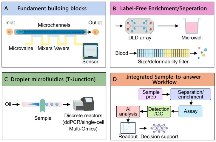
A useful way to picture a microfluidic chip is as a miniature plumbing system for biology. Instead of pipes carrying liters of water, the chip contains tiny channels that steer droplets or thin streams of blood, plasma, or chemical reagents through specific testing steps.
That miniaturization brings practical benefits. Compared with conventional centralized laboratory testing, these devices can be portable, fast, and highly integrated, meaning several steps that once required separate instruments can happen on one chip with less hands-on work.
Why point-of-care testing benefits most
The strongest case for microfluidics is in point-of-care testing, which means diagnostic testing performed near the patient rather than in a distant lab. In resource-limited settings especially, the ability to identify disease quickly can reduce delays in treatment and lower the burden of follow-up visits that some patients may not be able to make.
The source material notes that microfluidic POCT devices are now being deployed in a wide range of settings, including pre-hospital emergency care, community clinics, and home environments. That breadth matters because each setting has different constraints, from limited staff and electricity to the need for simple workflows that non-specialists can use.
Moving into cancer marker detection
One of the more technically ambitious areas is the analysis of acellular tumor markers, which are cancer-related traces found in body fluids rather than in whole tumor cells. Two important examples are circulating tumor DNA, or ctDNA, which consists of fragments of tumor-derived genetic material in blood, and exosomes, tiny vesicles released by cells that can carry proteins and nucleic acids.
These markers are valuable because they can support so-called liquid biopsy approaches, where a blood sample may reveal information about a tumor without an invasive tissue procedure. Microfluidic platforms have made progress here by combining sample preparation with sensitive molecular testing on the same chip, reducing manual transfer steps that often slow testing and introduce error.
What integration looks like on the chip
The review highlights how newer systems do more than detect a target at the end of a workflow. They can first separate useful material from complex samples like whole blood, then enrich the target, and finally run a molecular readout, all within one connected device.
One example described in the source is the use of droplet digital polymerase chain reaction, or ddPCR, microfluidics to quantify ctDNA. PCR is a method for copying genetic material so it can be measured, and the droplet digital version divides a sample into many tiny partitions, a bit like checking thousands of miniature reaction tubes at once, which improves sensitivity and allows absolute counting rather than rough estimation.
The source also points to a finger-powered microfluidic chip that rapidly processes whole blood and uses a surface-enhanced Raman scattering, or SERS, assay. Raman scattering reads how light interacts with molecules, and the surface-enhanced version boosts that signal using specially designed surfaces, making it easier to detect faint molecular traces from very small samples.
A modular, LEGO-like redesign
Not all progress is about chemistry or sensitivity. Some of the most interesting work changes how chips are built in the first place through modular microfluidic design, where interchangeable units can be assembled into different fluidic circuits as needed.
The easiest analogy is LEGO bricks: instead of fabricating a custom device from scratch every time, researchers can snap together standard modules for mixing, filtration, or cell sorting. According to the source, injection-molded modules with standardized connectors have been developed to make this possible, which could reduce prototyping time and help solve a long-standing problem in microfluidics: lack of standardization across devices.
The review specifically mentions researchers at MIT who demonstrated a plug-and-play platform made of LEGO-like blocks. By joining those blocks in different sequences, they could perform functions such as blood cell separation and chemical gradient generation, showing how one hardware kit might support many lab tasks rather than a single fixed assay.
Where artificial intelligence fits in
The article frames artificial intelligence integration as an important next step for laboratory microfluidics. In plain terms, AI can act as the chip's pattern reader, helping interpret noisy, high-dimensional outputs that may be difficult for a human operator to analyze quickly or consistently.
That could be especially useful when a chip collects complex optical, molecular, or imaging signals. Instead of simply producing a yes-or-no result, future systems may combine microfluidic sample handling with software that classifies patterns, estimates risk, or flags borderline cases for follow-up testing.
AI also fits the practical goals of point-of-care testing. If a device is going to be used in a community clinic or at home, automated interpretation can reduce dependence on highly trained personnel and make results more consistent across settings.
Why This Matters
The bigger story is not just that microfluidic chips are getting smaller or smarter. It is that laboratory medicine is shifting toward tools that compress multiple steps into one workflow, bringing sophisticated analysis closer to where patients are actually seen.
That matters for speed, but also for equity. A portable, cost-effective test that works outside a major hospital can widen access to diagnostics, especially in places where centralized lab infrastructure is limited or where delays in transport and processing can blunt the value of a result.
The cancer-testing examples show another reason this field is worth watching: microfluidics is moving into applications where sample volumes are tiny, targets are rare, and every handling step can affect accuracy. If chips can reliably isolate and detect materials like ctDNA or exosomes with minimal manual intervention, they may help make advanced molecular testing more routine.
There are still hurdles, including standardization, validation in real-world clinical workflows, and the challenge of integrating AI in ways clinicians trust. But the direction is clear: lab-on-a-chip systems are evolving from clever miniaturized gadgets into more complete diagnostic platforms, with modular design and software intelligence likely to shape the next phase of adoption.