Liquid biopsy aims to find cancer by reading traces it leaves in blood and other body fluids, and microfluidic technology is becoming one of the most useful tools for that job. In this review, researchers describe how tiny chip-based devices can isolate and analyze circulating tumor cells, cell-free nucleic acids, exosomes, and proteins using far smaller samples than many standard lab methods require. The basic idea is simple: instead of sending a tube of blood through several bulky instruments, a microfluidic chip guides minute volumes through narrow channels that sort, trap, and test rare cancer signals with high precision. That matters because the most valuable biomarkers in liquid biopsy are often extremely scarce, like a few mutant DNA fragments or a handful of tumor cells hidden among millions of normal cells. The reviewed studies show that these chips can sometimes match the accuracy of polymerase chain reaction, or PCR, while cutting sample preparation, analysis time, and cost. They also point to more specialized advances, including droplet-based digital PCR for oncogene mutations and electrochemical sensors that detect mutant nucleic acids directly from serum. At the same time, the review is careful about limitations, especially around separating target cells cleanly from healthy blood cells and proving performance in real patient samples. Taken together, the article presents microfluidics not as a single test, but as a growing toolkit that could make cancer monitoring faster, less invasive, and more practical in clinical care.
Why microfluidics fits liquid biopsy
Think of a microfluidic chip like a highly organized road system for droplets and cells. Instead of letting everything mix in a large test tube, the chip directs tiny amounts of fluid through precise paths so rare targets can be captured or measured before they get lost in the crowd.
That level of control is particularly useful in cancer liquid biopsy because the material doctors want to study is often vanishingly rare. A blood sample may contain a small number of circulating tumor cells, or CTCs, along with fragments of tumor DNA and RNA called cell-free nucleic acids, mixed into a huge background of normal blood components.
Detecting mutated DNA and RNA on-chip
One major theme in the review is the use of microfluidic devices to detect cancer-linked mutations in cell-free nucleic acids. These are snippets of genetic material shed by tumors into body fluids, and they can carry mutations in genes such as KRAS, an oncogene often altered in cancer.
The review highlights work by Pekin and colleagues, who built a microfluidic chip for microdroplet-based digital PCR. In that setup, genomic DNA and mutation-specific TaqMan probes were partitioned into droplets, turning each droplet into a tiny reaction chamber; green and red fluorescence then revealed the relative amounts of mutant and normal gene sequences. According to the review, the system was sensitive enough to detect cell-free DNA in blood, stool, and lymph samples while showing strong specificity for oncogene mutation detection.
Electrochemical sensing without amplification
Another advance came from Das and colleagues, who reported what the review describes as the first electrochemical approach for direct detection of mutated cell-free nucleic acids from serum. Their chip used an electrochemical clamp assay, a method that relies on molecules designed to bind specific sequences while clamp molecules reduce cross-reactivity, meaning fewer false matches to similar but incorrect targets.
An everyday analogy is a lock that accepts one key while actively blocking keys that almost fit. On the chip, that selectivity came from a peptide nucleic acid, or PNA, modified microsensor, which allowed rapid and sensitive detection of cancer-related mutations from as little as 5 femtograms of isolated RNA without enzymatic amplification. The authors reported accurate detection of mutated sequences in serum samples from lung cancer and melanoma patients.
Why skipping purification can matter
One practical advantage emphasized in the review is that some chip-based methods can work with unpurified serum. In ordinary workflows, purification steps often add time, cost, and opportunities to lose fragile material, especially when the starting amount is already tiny.
The reviewed chip approach was reported to achieve accuracy comparable to PCR while using less sample, shortening analysis time, and lowering cost per test. For a clinical lab, those details are not minor conveniences; they can determine whether a promising technology stays in a research paper or becomes realistic for routine monitoring.
Sorting and identifying tumor cells in blood
Microfluidics is not just for DNA and RNA. The review also covers efforts to detect whole cancer cells in peripheral blood, which can provide different information because intact cells preserve morphology, protein expression, and other features that free-floating DNA cannot capture.
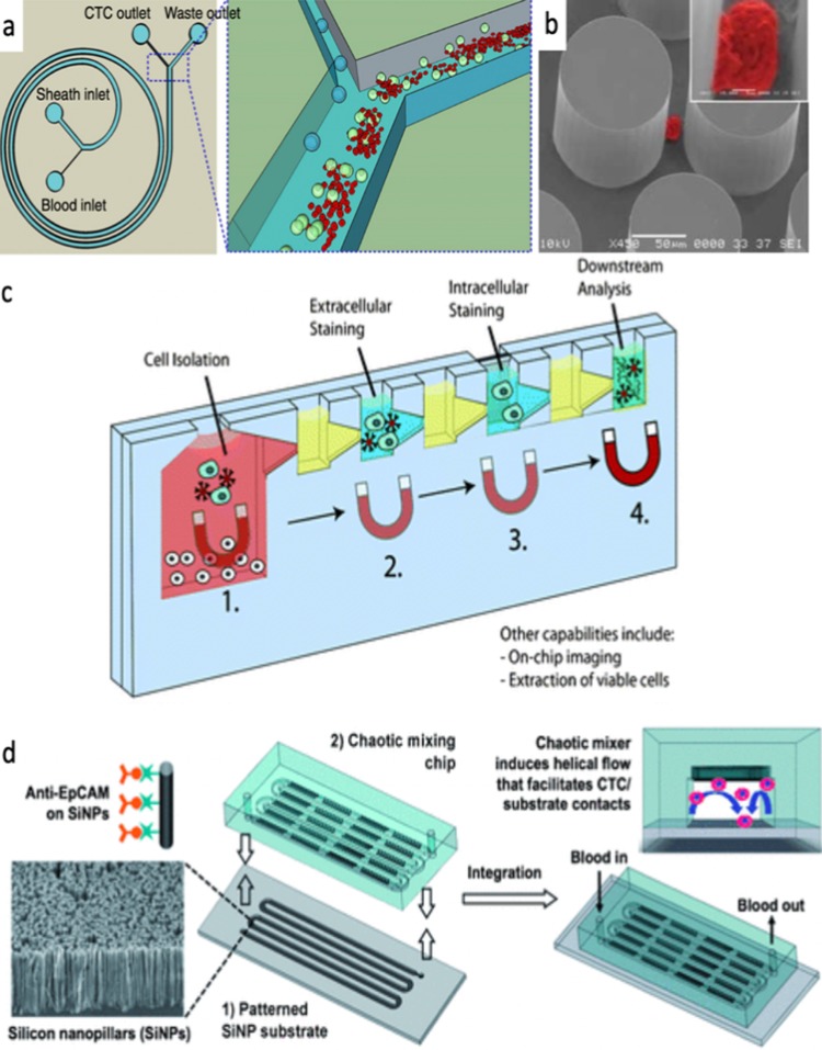
In one example involving suspended breast cancer cells, red blood cells were first removed continuously using a magnetophoretic microseparator, a device that uses magnetic forces to sort cells. A silicon chip downstream then captured and analyzed the remaining cells using electrical impedance spectroscopy, or EIS, which measures how cells respond to an applied electrical signal.
What impedance can and cannot tell you
EIS works a bit like tapping different objects and listening to how they resonate. Because cells vary in size, membrane structure, and internal composition, they produce distinct electrical signatures that can help distinguish one cell type from another.
In the breast cancer study described in the review, the chip differentiated among the human breast cancer cell lines MCF-7, MDA-MB-231, and MDA-MB-435, and it separated them electrically from the normal breast cell line MCF-10A. But the review also points out a key weakness: even if the electrical readout cleanly separates cancer cells from normal ones, it remained unclear how efficiently healthy but unwanted cells, especially white blood cells, could be removed from the sample before analysis.
Why This Matters
The promise of liquid biopsy is straightforward: track cancer with a blood draw instead of repeatedly relying on invasive tissue biopsies. Microfluidic systems could help that promise become routine by shrinking complex laboratory procedures into compact devices that need less sample, less hands-on work, and potentially less money.
That could be useful across the cancer timeline. Earlier detection of tumor-derived mutations, quicker monitoring of treatment response, and repeated sampling to watch resistance emerge are all easier to imagine when tests can be run rapidly on small volumes and with minimal preparation.
The remaining gap between prototypes and practice
The review also makes clear that technical elegance is not enough. A chip that performs well with cell lines or carefully prepared samples still has to prove it can handle the messiness of real clinical specimens, where rare targets appear in unpredictable amounts and healthy cells or molecules can interfere with the signal.
Standardization is another hurdle. To move from promising prototypes to everyday diagnostics, researchers will need reproducible manufacturing, robust validation across larger patient groups, and workflows that integrate smoothly with hospital laboratories and existing pathology practices.
What comes next
The broader direction is encouraging: microfluidic liquid biopsy is evolving from simple enrichment tools into integrated systems that can isolate, detect, and quantify multiple cancer biomarkers on the same platform. If future studies can show reliable performance in real-world samples and clarify where each chip works best, these devices could become a practical bridge between sophisticated molecular biology and the ordinary clinic visit.