Researchers at the University of Sydney have built a transparent blood vessel-on-a-chip, a tiny lab device designed to imitate what happens inside human arteries as heart disease begins to develop. The chip recreates two of the major forces involved in early vascular injury: unusually high blood flow stress and inflammation, both of which can damage the cells lining blood vessels. Because the device is transparent, scientists can watch these changes unfold under a microscope in far greater detail than is possible in animal studies. That matters because the earliest stages of heart disease are difficult to observe directly, even though they help determine where dangerous blockages later form. The team says the new platform could improve how researchers study why plaques and damage appear in specific parts of blood vessels rather than others. It could also become a more realistic way to test new drugs before moving into clinical trials, potentially reducing some dependence on animal experiments. More broadly, the work points toward a future in which organ-like chips are used to model disease in the lab with much finer control and visibility. If that promise holds up, devices like this could become important tools for both cardiovascular research and drug development.
A clearer window into heart disease
The chip was developed by a University of Sydney team led in part by Associate Professor Anna Waterhouse of the Charles Perkins Centre and the University of Sydney Nano Institute. Their goal was to create a system that better captures the physical conditions inside the coronary arteries, the vessels that supply blood to the heart muscle.
Heart disease does not begin with a sudden blockage. It usually starts with subtle damage to the inner lining of blood vessels, known as the endothelium, which can be triggered by abnormal flow patterns and inflammatory signals. Over time, those damaged regions can become hotspots where fatty deposits and other materials accumulate.
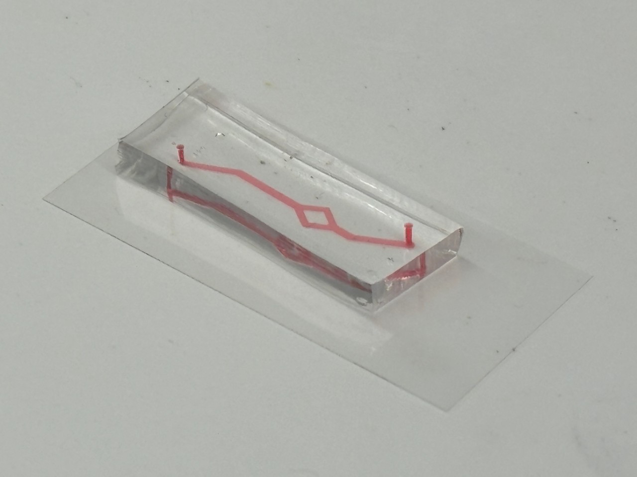
Why transparency changes the experiment
The standout feature of the device is its transparency. That may sound simple, but it gives researchers a major advantage: they can directly image living cells inside the chip and map where damage appears as the experiment runs.
According to Waterhouse, the team used the chip to mimic conditions found in coronary arteries and then examined the resulting cell damage with a microscope. The damaged zones matched locations similar to where blockages are seen in patients with heart disease, suggesting the model may capture meaningful aspects of how disease starts in the body.
What the chip is designed to mimic
In essence, a vessel-on-a-chip is a type of microfluidic device, meaning it uses tiny channels to control the movement of fluids such as blood-like solutions. In this case, those channels are lined with cells so the system behaves more like a miniature blood vessel than a simple plastic tube.
The Sydney team specifically focused on damage caused by high blood flow and inflammation. Those are important because blood does not move evenly through every part of the vascular system; bends, branches, and narrowing can create mechanical stresses that affect how vessel-lining cells behave, sometimes making them more vulnerable to injury.
An advance in the chip's surface engineering
The researchers also highlighted a new way of preparing the chip's surface so that coatings and biomolecules attach more firmly. Biomolecules are the proteins and other biological compounds that cells need in order to stick, grow, and behave normally on an artificial surface.
That step is more important than it sounds. If cells do not attach properly, the device cannot realistically mimic blood flow or vessel biology, so improving the bonding chemistry makes the platform more stable and useful for experiments that aim to model disease.
Why animal models have limits here
One reason the work stands out is that it addresses a basic problem in cardiovascular research: it is hard to see early vessel damage inside a living animal at microscopic resolution. Even if an animal model captures some aspects of disease, researchers often cannot directly watch individual cell responses in the way they can in a transparent chip.
Waterhouse made that contrast explicit, noting that changes at this level of detail are not visible in intact blood vessels in living organisms. The chip therefore acts as both a model and an observation platform, letting scientists study not just outcomes but the sequence of events that leads to them.
Potential use in drug development
The team says the technology could help reduce animal testing for drugs aimed at treating heart disease. A better preclinical model, meaning a test system used before human trials, can give researchers earlier clues about whether a therapy protects vessel-lining cells, reduces inflammation, or prevents the kinds of damage associated with later blockages.
That does not mean chips like this will replace animal studies or clinical trials overnight. But they can serve as an intermediate step that is more human-relevant than simple cell cultures and more observable than whole-animal models, which could improve screening decisions and save time in the development pipeline.
Beyond blood vessels
The researchers say the findings from their studies could support broader use of the chip in the biomedical field to model human organs and diseases. In other words, the same design principles could be adapted to recreate parts of the body where fluid flow, cell interactions, and tissue damage matter.
The team is already looking ahead to devices that simulate more complex interactions within organs, with the aim of reproducing later and more severe disease stages. Waterhouse said that could include advanced heart disease and even cancers, where the local tissue environment strongly shapes how disease develops and how treatments work.
Why This Matters
This work fits into a growing movement toward organ-on-a-chip systems, small engineered devices that mimic the behavior of living tissues. What makes the Sydney advance notable is the combination of disease realism and direct visibility: the chip does not just imitate a vessel, it allows researchers to watch damage emerge in places that resemble real-world disease patterns.
That could be valuable for basic biology, because scientists still do not fully understand why some parts of arteries are especially prone to disease. It could also matter for medicine, since a more accurate model of early vascular injury may help researchers evaluate drugs, compare treatment strategies, and eventually design more personalized approaches to preventing heart disease.
What comes next
The next challenge will be showing that the platform remains reliable as experiments become more complex. To be truly useful, future versions will need to incorporate additional cell types, more realistic immune responses, and longer-term disease progression while still preserving the imaging advantages that make the chip so powerful.
Even so, the new transparent vessel-on-a-chip is a strong example of how engineering can open up parts of human biology that were previously hard to study. By turning the earliest steps of vascular disease into something scientists can directly observe, the device offers a promising new way to understand, and eventually better treat, one of the world's leading causes of death.