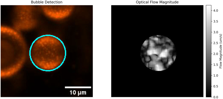
Researchers studying microbubbles—tiny gas-filled spheres used in ultrasound imaging and drug delivery—are getting a clearer picture of why bubbles that look identical can behave very differently. In this study, the authors focused on the bubble’s outer lipid shell, a thin fatty coating that acts a bit like the skin of a soap bubble but with much more complex chemistry. That shell does not always form as a perfectly even layer. Instead, different lipids can cluster into patches, and those patches appear to change how a bubble expands, compresses, survives, and scatters sound. To probe that problem, the team used fluorescent membrane probes, labeled lipids, and high-resolution confocal microscopy to watch how the shell organized itself during bubble formation and during changes in bubble volume. They also introduced a simple heat-based preparation approach they call the cocktail method, designed to make microbubbles look more uniform and less obviously divided into domains. Even with that treatment, the study found that the chemical nature of key lipids still pushed the shell toward some degree of phase separation. The result is a more nuanced view of microbubble design: surface uniformity matters, but some built-in material properties may keep complete homogeneity out of reach.
Why microbubble shells matter
Microbubbles are already important tools in medicine because they interact strongly with ultrasound waves. Clinicians can use them as contrast agents to make blood flow and tissue structures easier to see, and researchers are exploring them as carriers that help deliver drugs to precise places in the body.
But for either job, consistency matters. If two bubbles from the same batch respond differently to the same ultrasound pulse, the result can be noisier images, misleading signal patterns, or less precise targeting during bubble-assisted therapy.
A hidden source of variability
This study starts from a problem that has become increasingly visible in ultrasound research: even monodisperse microbubbles, meaning bubbles selected to be very similar in size, can still show very different acoustic responses. Size alone is not enough to explain that spread in behavior.
The authors point to the shell as a likely reason. Think of two tennis balls with the same diameter but different surface materials—one smooth and one partly stiffened with tape. They may look alike from far away, yet bounce differently. In the same way, microbubbles with the same diameter may still react differently if their shells vary in organization, stiffness, or viscosity.
Watching the shell form in real time
To study that organization, the researchers combined several imaging tools that let them distinguish where different lipids were located and how those lipids behaved. Using fluorescent labels and membrane-sensitive probes, they tracked surface dynamics, phase behavior, and micro-viscosity, which is a local measure of how freely molecules can move within tiny regions of the shell.
High-resolution confocal microscopy gave them a close-up view of the bubble surface during two critical moments: when the shell first formed and when the bubble changed volume. Those expansion and compression cycles matter because ultrasound repeatedly squeezes and stretches a microbubble. A shell that reorganizes under stress may also change its acoustic signature.
The “cocktail method”
The paper’s practical contribution is the cocktail method, described as a straightforward thermal procedure intended to produce seemingly domainless microbubbles. In simple terms, the idea is to use heat to encourage the shell components to mix more evenly, reducing visible patchiness on the bubble surface.
That matters because visible domains often signal phase separation, where different lipids prefer different local neighborhoods rather than forming one smooth blended layer. If a more homogeneous shell also behaves more predictably under ultrasound, then preparation method becomes just as important as composition.
What the researchers found
The study identified distinct behaviors among individual lipid components during shell formation. That means the shell is not just a passive coating assembled all at once; its ingredients contribute differently as the structure takes shape.
The authors also provide a qualitative picture of viscosity in specific lipid phases during compression and expansion. In everyday terms, some regions of the shell act more like a flexible oil film, while others behave more like a crowded, thicker layer. Those local differences can affect shell elasticity, the tendency of the bubble surface to resist deformation and then recover its shape.
Most importantly, the paper links lipid organization to acoustic behavior. When the shell is more heterogeneous, its mechanical response under ultrasound may become less uniform, helping explain why bubbles that seem similar can emit different signals.
Why full uniformity may be hard to achieve
One of the study’s central findings is that the physicochemical properties of two lipids, DSPC and DSPE-PEG5000, appear to drive an unavoidable degree of phase separation. DSPC, or distearoylphosphatidylcholine, is a saturated phospholipid often used to build relatively rigid shells. DSPE-PEG5000 is a lipid linked to polyethylene glycol, a polymer that can change how molecules pack and interact at the surface.
Even after thermal quenching—rapid cooling intended to lock in a more mixed arrangement—the tendency toward separation persisted. That suggests the shell is governed not just by processing history but by the built-in preferences of its ingredients. In other words, the bubble can be coached toward uniformity, but the chemistry still has the final say.
Why This Matters
This is an important insight for anyone trying to build better ultrasound agents. If shell heterogeneity alters stability, elasticity, and sound scattering, then microbubble performance cannot be optimized by controlling size alone. Researchers may need to think more carefully about lipid selection, shell architecture, and manufacturing conditions together.
The findings are especially relevant for high-resolution ultrasound imaging and for applications such as microbubble-assisted drug delivery, where localization accuracy is critical. A bubble that responds unpredictably could introduce imaging artifacts or reduce the precision of energy delivery at a target site.
Just as importantly, the work highlights a design constraint. Some phase separation may be intrinsic to widely used lipid mixtures, which means the goal may shift from eliminating heterogeneity entirely to understanding it well enough to manage and exploit it.
What comes next
The study does not present microbubble heterogeneity as a minor manufacturing flaw. It frames it as a fundamental materials problem that sits at the center of how these agents function. By tying shell organization to viscoelastic and acoustic behavior, the authors give the field a more detailed map of where variability comes from.
That map could guide the next generation of microbubbles toward more predictable imaging and therapy performance. Future work will likely test additional lipid combinations, connect microscopic shell patterns to measured acoustic outputs more directly, and look for formulations that balance stability with consistent ultrasound response. For a technology built on tiny bubbles, the message is surprisingly large: the fine structure of the shell may determine whether a microbubble is merely visible or truly reliable.