Source: Cell Reports Medicine, by Syeda Maheen Batool; Anudeep Yekula; Prerna Khanna; Tiffaney Hsia; Austin S Gamblin; Emil Ekanayake; Ana K Escobedo; Dong Gil You; Cesar M Castro; Hyungsoon Im; Tugba Kilic; Michelle Andrea Garlin; Johan Skog; Daniela M Dinulescu; Jonathan Dudley; Nishant Agrawal; Jordan Cheng; Fereidoun Abtin; Denise R Aberle; David Chia. AI-generated summary by biochip.com, published . Not independently reviewed.
A broad group of researchers working under the Liquid Biopsy Consortium is trying to answer a hard medical question: can cancer be found and tracked through a simple sample of blood or other body fluids, instead of surgery or repeated tissue biopsies? The consortium’s report lays out both the promise and the practical barriers facing that idea. It describes a fast-moving field built around liquid biopsy, an umbrella term for tests that look for traces of cancer such as circulating tumor DNA, whole tumor cells, extracellular vesicles, proteins, and small RNA molecules in fluids like plasma or pleural fluid. The attraction is obvious. A blood draw is less invasive, easier to repeat over time, and potentially better at capturing how a tumor changes than a single tissue sample taken from one place on one day. But the report is equally clear that this is not yet a one-size-fits-all answer: the rare signals are technically difficult to isolate, different cancers shed different markers, and study designs still need to show that these tools improve real-world outcomes. The consortium argues that progress will depend not just on better chips, sequencing, and imaging, but on carefully designed prospective studies that compare liquid biopsy methods with standard care. Its central message is that early cancer detection and monitoring through fluids is plausible and increasingly sophisticated, yet success will require rigorous validation, realistic expectations, and close collaboration between engineers, clinicians, industry, and the public.
A field built around faint signals
Liquid biopsy works a bit like trying to identify a ship by the wake it leaves behind. Instead of removing a piece of the tumor itself, researchers search body fluids for tiny biological traces that the tumor releases. Those traces can include ctDNA, or circulating tumor DNA, which is fragmented genetic material from cancer cells; CTCs, or circulating tumor cells, which are whole cells that have broken away from a tumor; and extracellular vesicles, small membrane-bound packages that cells use to send molecular cargo.
The challenge is that these signals are rare, especially in early-stage disease. A blood sample may contain only a handful of relevant cells or fragments among vast amounts of normal material. That is why the consortium emphasizes sensitivity, specificity, and careful matching of the right analyte to the right clinical question.
Many technologies, no single winner
The report surveys a long list of technologies now being used to chase these tumor signals. On the DNA side, researchers are using polymerase chain reaction methods such as ddPCR, or droplet digital PCR, and methylation-based PCR, alongside targeted sequencing, deep sequencing, and larger next-generation sequencing panels. In plain terms, these methods either look for known mutations with high precision or scan broader stretches of genetic material for patterns associated with cancer.
Other tools focus on cells and particles rather than DNA fragments. These include microfluidic systems, density gradient centrifugation, imaging flow cytometry, mass spectrometry, enzyme-linked immunosorbent assay, and microfluidic nuclear magnetic resonance. Think of these as different kinds of sieves, scanners, and chemical readers: some sort by size or density, some identify cells by their appearance, and some detect proteins or other molecules that may reveal a tumor’s presence.
How chips and automation fit in
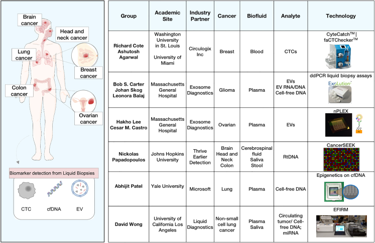
One example highlighted in the source material involves fibroblasts from early-stage breast cancer patients. Fibroblasts are connective-tissue cells, and in this context researchers are interested in rare cells associated with the disease that can be recovered from blood or other biofluids. To isolate them, the group uses a microfilter technology called CyteCatch together with an automated fluidic platform called faCTChecker.
Microfilters work much like a very precise kitchen strainer, except the goal is to separate unusually rare cells from everything else flowing past. After capture, the cells are analyzed on-chip through image analysis, and those images are being used as training data for deep-learning systems intended to automate future identification. The consortium notes industry partners including Circulogix for the microfilter platform and Google for the deep-learning component, showing how the field now spans hardware, software, and clinical research.
Different cancers call for different liquid biopsies
The report also makes a practical point that often gets lost in hype: there is no universal liquid biopsy marker that works equally well for every tumor type. In breast cancer, the source lists work using plasma-based ctDNA, CTCs, and cell-free microRNA, or small RNA molecules that can regulate gene activity. The associated methods include BEAMing, ddPCR, quantitative reverse-transcription PCR, TEC-seq, personalized ultra-deep sequencing, and large sequencing panels, along with platforms such as the nanotube-CTC chip and CTC-iChip.
For lung cancer, researchers are drawing from plasma and pleural fluid and studying extracellular vesicles, CTCs, ctDNA, and microRNA. The toolkit there includes PCR and qRT-PCR, ARMS-PCR, and CAPP-seq, among others. The pattern is important: cancers differ in where they shed material, how much they shed, and which biomarkers are most informative, so test design has to be tailored rather than assumed.
The hardest part is proving clinical value
Building a sensitive assay is only the first hurdle. The consortium argues that the next big step is designing prospective studies that show where liquid biopsy truly helps patients. That means asking specific questions: can these tests detect cancer early enough to shift diagnosis toward less advanced stages, reduce unnecessary invasive procedures, improve detection efficiency, or add information that standard care misses?
Those questions matter because analytical performance alone does not guarantee benefit in the clinic. A test might detect something unusual, yet still lead to false alarms, overtesting, or uncertainty if doctors do not know how to act on the result. The report pushes for head-to-head comparisons with existing standards of care so the field can move beyond technical feasibility toward practical evidence.
Why This Matters
If liquid biopsy reaches its goals, it could change how cancer care unfolds across the entire disease timeline. Earlier detection could mean more patients are diagnosed when treatment is simpler and outcomes are better. Repeated blood-based monitoring could also help doctors track treatment response, spot relapse sooner, and capture tumor evolution without repeated invasive sampling.
At the same time, the consortium is careful not to oversell the technology. It argues that tests must be cost-effective, rigorous, and complementary to the existing marketplace rather than treated as automatic replacements for established diagnostics. Just as important, researchers and companies need to work with health-care professionals, including primary care physicians, and with the public to manage expectations while the technologies are still being refined.
What comes next
The overall picture is one of a field that has matured technically but is still sorting out how best to deliver useful answers in the clinic. The tools now range from microfluidic chips and imaging systems to ultra-deep sequencing and machine learning, yet the real measure of success will be whether they improve decisions and outcomes for patients. The consortium’s report suggests that the future of liquid biopsy will not depend on a single dazzling platform, but on matching the right marker, method, and study design to each cancer and each clinical use. If that happens, blood and other biofluids could become not just convenient samples, but powerful windows into cancer biology over time.