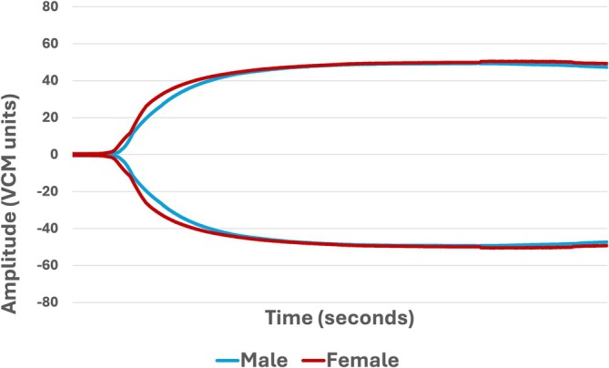
A small study of bonobos suggests that their blood may clot faster and more firmly than standard veterinary benchmarks would predict, a pattern that could matter when these endangered apes are treated for heart disease, stroke risk, or cancer. Researchers evaluated 12 bonobos during anesthetized health exams and tested their blood with the VCM Vet, a point-of-care viscoelastic coagulation monitor that tracks how a clot starts, strengthens, and breaks down over time. They also compared those readings with thromboelastography, or TEG, a more established method for measuring the same broad features of clotting. The central finding was that the bonobos appeared hypercoagulable, meaning their blood tended to form stronger or faster clots, when their results were compared with canine reference intervals. That pattern matches earlier observations in chimpanzees and hints that great apes may have species-specific clotting traits that do not fit neatly into dog-based veterinary standards. The study also found an important exception: bonobos receiving medication for hypertension, or high blood pressure, appeared more hypocoagulable, meaning less prone to clot, than normotensive animals. For zoo and wildlife veterinarians, the work offers early baseline data for a rare species and a practical look at whether a bedside-style analyzer can help guide clinical decisions when time and sample volume are limited.
Why the researchers looked at bonobo clotting
Bonobos are endangered and relatively uncommon in managed care, which means veterinarians have limited species-specific data to rely on. That gap becomes especially important because prothrombotic states, where the body is biased toward clotting, and hypofibrinolytic states, where clots do not break down easily, are seen alongside conditions such as cardiovascular disease, ischemic stroke, and neoplasia.
A useful everyday analogy is wet cement. Traditional clotting tests can tell you when the cement starts to set, but viscoelastic tests show the whole process: when it first thickens, how solid it becomes, and whether it later softens or cracks apart. For clinicians, that fuller picture can be more informative than a single snapshot.
How the study was done
The researchers studied 12 bonobos from a single institution during anesthetized examinations. Using blood collected during those procedures, they ran samples on a VCM Vet analyzer, which produced results within one hour.
They then compared the VCM Vet findings with thromboelastography and with conventional coagulation testing. That side-by-side approach matters because newer point-of-care devices are only useful in practice if their readouts line up reasonably well with methods veterinarians already trust.
What the VCM Vet measured
The VCM Vet reports several parameters that map onto different stages of clotting. In this study, median values showed a clot time of 300 seconds, with an interquartile range of 265 to 332 seconds, and a clot formation time of 97 seconds, with an interquartile range of 82 to 107 seconds.
The analyzer also measured the alpha angle, reported here as 65 degrees with an interquartile range of 63 to 69. In plain language, that angle reflects how quickly the clot gains structure after it begins to form, much like watching how fast gelatin firms up once it starts setting.
How strong the clots were
To estimate clot strength, the team looked at maximum clot formation and clot amplitude over time. Median maximum clot formation was 50 units, with an interquartile range of 47 to 52; amplitude at 10 minutes was 40 units, with an interquartile range of 35 to 44; and amplitude at 20 minutes was 47 units, with an interquartile range of 43 to 51.
Those values suggest that once clotting started, the clots built substantial firmness. In viscoelastic testing, stronger traces usually point to more robust clot structure, which can reflect the combined effects of platelets, fibrin formation, and other parts of the coagulation system working together.
What happened to clot breakdown
The study also tracked clot lysis, the process by which a formed clot is gradually dissolved. The median lysis index at 30 minutes was 99.97%, with an interquartile range of 99.95 to 100.00, and at 45 minutes it was 98.10%, with an interquartile range of 97.50 to 99.20.
Those numbers indicate very little clot breakdown over the measured period. That is consistent with a hypofibrinolytic pattern, meaning the body is not rapidly clearing clots once they form. In the right clinical context, that can contribute to a higher-thrombosis picture.
How bonobos compared with existing references
The researchers note that bonobos were hypercoagulable when compared with canine reference intervals. That does not automatically mean the animals were abnormal in a species-specific sense; it may instead show the limits of using dog-based standards for a great ape.
That distinction is crucial. If clinicians interpret bonobo bloodwork through the wrong species lens, they could mistake a normal bonobo pattern for disease, or miss a true problem because the comparison group is poorly matched. The similarity to chimpanzee findings strengthens the case that this may be a broader ape-related trait.
A notable signal in bonobos with hypertension treatment
One of the study's most clinically interesting observations was that bonobos medicated for hypertension appeared hypocoagulable compared with normotensive individuals. In other words, within this already small group, animals being treated for high blood pressure showed a relative shift toward weaker or slower clotting.
The source does not establish why that difference appeared, and the small sample size means it should be interpreted cautiously. Even so, it raises practical questions for veterinarians about how cardiovascular disease, its treatment, or both might influence coagulation in bonobos.
Why This Matters
For rare species, baseline physiology is not a luxury; it is part of safe medicine. When a bonobo needs anesthesia, surgery, or evaluation for cardiovascular or neurologic disease, veterinarians need some way to estimate bleeding risk and clotting risk without waiting for lengthy laboratory processing or relying entirely on other species.
This study suggests that the VCM Vet could be useful in that role because it delivers a broad coagulation profile within an hour and can be compared with TEG and routine coagulation tests. Just as important, the findings underline that bonobos may have their own characteristic clotting profile, one that could affect how clinicians interpret results, monitor disease, and manage treatment. The next step will be larger studies across more institutions to separate normal species biology from the effects of age, disease, medication, and captivity-related health conditions.