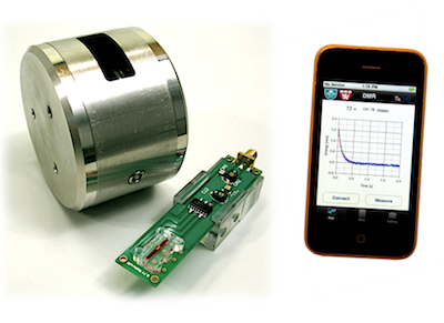
A research team at Massachusetts General Hospital and Harvard Medical School developed a compact cancer-testing system designed to do something pathology often cannot do quickly: identify whether suspicious cells are likely cancerous in about an hour. The platform combines a small microchip, magnetic nanoparticles, and a handheld NMR reader—short for nuclear magnetic resonance, a technique that detects molecular changes using magnetic signals. Instead of waiting for tissue to be processed in a lab over many hours or even days, doctors could potentially analyze a fine-needle sample close to the patient’s bedside. The group also linked the system to a smartphone-style interface, making the results easier to read and transmit. In early studies, the device appeared highly accurate at detecting tumor cells from small biopsy samples. The bigger idea was not just speed, but bringing sophisticated cancer diagnostics out of specialized labs and into routine clinical care. If that promise holds up in larger trials, tools like this could make cancer diagnosis faster, cheaper, and more accessible.
A lab technique shrunk to bedside size
The work came from the MGH Center for Systems Biology, where researchers were trying to miniaturize one of medicine’s more cumbersome tasks: analyzing a biopsy. A biopsy usually means removing a small amount of tissue so pathologists can examine it for signs of disease, especially cancer.
Traditional pathology is powerful, but it takes time. Samples often need to be preserved, sliced into thin sections, stained with dyes, and reviewed by specialists, which can delay decisions about surgery or treatment.
How the system works
The new setup uses tiny magnetic particles that bind to specific proteins on the surface of cells. Those proteins act like biological name tags, helping distinguish potentially malignant cells from normal ones.
Once the particles attach, a miniaturized NMR device reads the magnetic signal they produce. In plain terms, the stronger or more specific the signal, the more confidence doctors may have that the sampled cells carry the molecular features associated with cancer.
This is different from imaging tools like MRI, even though both rely on magnetic principles. Here, the goal is not to create a picture of the body, but to detect molecular markers in a very small sample of cells.
Why speed matters in cancer care
When a patient has a suspicious lump or lesion, waiting for an answer can be agonizing. Fast results could help clinicians decide sooner whether they need another biopsy, more imaging, immediate surgery, or a different treatment plan.
Speed also matters during procedures. Surgeons and interventional radiologists often need to know whether they collected enough material from a needle biopsy; a rapid bedside test could reduce repeat sampling and help confirm that the sample is actually useful.
The smartphone connection
Part of the public excitement around the project came from its phone-linked design. Rather than requiring a large, fixed workstation, the device could send results to a mobile interface, making it look more like modern consumer electronics than traditional hospital hardware.
That matters because usability is often the hidden barrier in medical technology. A diagnostic tool that is compact, portable, and easy to interpret stands a better chance of being used in busy clinics, outpatient centers, and eventually lower-resource settings.
What the early results suggested
According to reports about the study, the researchers tested the system on samples from patients with suspected tumors and found that it could identify cancer-associated cells with impressive accuracy. The device was also able to work with very small amounts of material, which is important because fine-needle biopsies do not always yield much tissue.
The team reportedly looked for multiple biomarkers rather than relying on a single molecular clue. That is a smart strategy, because cancer is not one disease; tumors vary widely, and using several markers can improve the odds of catching clinically meaningful differences.
What this could change in practice
If validated more broadly, a rapid microchip-based test could reshape how doctors move from suspicion to diagnosis. Instead of sending every specimen through a slower centralized workflow, some first-pass answers could be generated near the point of care, meaning where the patient is actually being seen.
That does not mean pathologists become unnecessary. More likely, tools like this would serve as a complement: a fast molecular screening method that helps triage samples, guide next steps, and focus traditional pathology where it is most needed.
The limits and open questions
Promising early results are not the same as routine clinical use. Any cancer diagnostic has to prove itself across many tumor types, many patient populations, and many real-world settings before hospitals can trust it for major decisions.
Researchers also need to show that the test is not just accurate in a controlled study, but reliable when used by ordinary clinical staff. Questions about cost, regulation, reimbursement, and manufacturing consistency can be just as important as the underlying science.
Why This Matters
This story matters because it captures a broader shift in medicine: moving complex diagnostics from centralized labs into smaller, faster, more connected devices. Cancer care in particular depends on timely answers, and every hour or day saved can reduce anxiety, speed treatment, and sometimes improve outcomes.
It also shows how biochip technologies can bridge disciplines. Here, microfabrication, nanotechnology, molecular biology, and mobile computing all work together to make a test that is not only scientifically clever but potentially practical.
The long-term vision is bigger than one cancer test. If researchers can keep shrinking advanced laboratory methods into portable systems, the same basic approach could eventually help diagnose infections, monitor treatment response, or characterize many diseases from tiny samples in near real time.