Source: WPI, by Not specified, Worcester Polytechnic Institute (December 15, 2016). AI-generated summary by biochip.com, published . Not independently reviewed.
Key takeaways
- WPI's nanotube-antibody chip captured EpCam- and Her2-positive breast cancer cells from blood samples spiked with target cells.
- The team used 170 wells and less than 0.85 milliliters of blood, reporting capture efficiency of 62 to 100 percent.
- The reported experiment used spiked blood samples, not clinical validation in patients with cancer.
Mechanical engineers at Worcester Polytechnic Institute developed a liquid biopsy chip designed to capture metastatic cancer cells from a small blood sample. The device uses antibody-coated carbon nanotubes in tiny wells, rather than the flowing-liquid format used by many microfluidic cancer-cell capture systems. In laboratory tests using blood spiked with breast cancer cells, the chip captured cells bearing two metastatic breast cancer markers, EpCam and Her2. WPI reported capture efficiencies ranging from 62 to 100 percent while using less than 0.85 milliliters of blood across 170 wells. The platform also aims to catch exosomes, tiny cancer-associated structures that can carry the same surface markers as tumor cells. That combination could eventually help clinicians detect signs of metastasis, the spread of cancer from its original site to distant organs, through a straightforward blood test. The research, led by Balaji Panchapakesan at WPI's Small Systems Laboratory, appeared in the journal Nanotechnology. Although the reported work was performed with spiked samples rather than a clinical patient-validation study, it demonstrates a distinct static chip design for isolating rare cancer-related material from blood.
Finding a Needle in a Blood Sample
Circulating tumor cells are cancer cells that have entered the bloodstream. They matter because metastasis often begins when cells leave an original tumor, travel through the body, and establish new tumors elsewhere. Breast cancer cells in circulation, for example, may later take hold in bone, lung, or brain tissue.
Detecting those cells is difficult because they are vastly outnumbered by normal blood components. Panchapakesan compared the challenge to finding a needle in a haystack: a sample can contain billions of red blood cells and tens of thousands of white blood cells, alongside only a small number of tumor cells. A device that selectively captures the rare cells could give researchers and clinicians a way to study them without taking a tissue sample from a tumor.
A Static Well Instead of a Flowing Channel
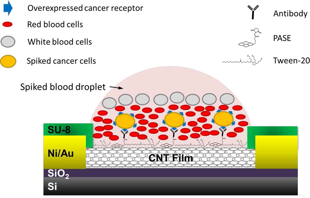
Many liquid biopsy systems use microfluidics, the control of very small volumes of fluid through narrow channels. In those devices, cells flow past antibodies anchored to a surface and must bind while moving. WPI's approach is different: blood sits in small wells, allowing cells to settle downward onto a capture surface.
Each chip element is about one-tenth of an inch, or 3 millimeters, across and contains a tiny well. At the well's bottom, antibodies are attached to an array of carbon nanotubes, hollow structures made from carbon that provide a nanoscale surface for the capture chemistry. Think of each well as a small fishing spot: the antibodies act as selective hooks, while the nanotube layer holds those hooks at the bottom where cells settle.
Matching Antibodies to Cell Markers
Antibodies are proteins that recognize particular molecular features. On this chip, each well can contain an antibody chosen to bind a cancer-cell surface marker, allowing the system to distinguish target cells from much more abundant blood cells. The concept also makes the array adaptable, because different wells can be prepared with different antibodies.
Panchapakesan's team used antibodies targeting EpCam and Her2, two markers associated with metastatic breast cancer cells in the reported experiment. When blood samples spiked with cells expressing those markers were placed on the chip, the device selectively captured the marked cells. The work involved WPI researchers as well as collaborators at the University of Louisville and Thomas Jefferson University, with postdoctoral researcher Farhad Khosravi serving as lead author of the journal paper.
What the Laboratory Test Measured
The researchers filled 170 wells using just under 0.3 fluid ounces, or 0.85 milliliters, of blood. Across the device, they captured from one to 1,000 cells and reported capture efficiency between 62 and 100 percent. Such measurements describe how well the chip recovered the target cells introduced into the test sample.
The chip also uses electrodes to sense electrical changes after cells bind to the capture surface. This readout gives the platform a way to detect captured cells directly on the device, rather than relying only on visual inspection. The reported study was titled “Static micro-array isolation, dynamic time series classification, capture and enumeration of spiked breast cancer cells in blood: the nanotube–CTC chip.”
Adding Exosomes to the Search
Beyond whole circulating tumor cells, the WPI team said its chip could capture exosomes. These are extremely small structures released by cells, including cancer cells, and they can carry molecular markers related to the cells that produced them. Panchapakesan described the relevant structures as 3 nanometers in size and said their small scale makes them difficult for flow-based devices to retain.
The proposed advantage is that a static well avoids the shear forces created by moving fluid. Shear force is the pulling and pushing stress generated when fluid flows across a surface, much like a strong current can tug at small objects in water. Panchapakesan said those forces can potentially destroy fragile exosomes in microfluidic systems, while the nanotube-antibody design may capture both tumor cells and exosomes on the same chip.
Why This Matters
A liquid biopsy seeks cancer-related information from blood rather than from a surgically collected tissue sample. For metastatic disease, that is especially appealing because tumor cells can circulate through the bloodstream and may represent cancer that is moving beyond its original site. The ability to test a small sample could also make repeated measurements more practical if the method is developed for clinical use.
Just as important, the chip is designed around specific markers. By populating different wells with an assortment of antibodies, the platform could be configured to seek several cancer-cell types from one blood sample. That marker-based approach could potentially help identify tumor characteristics relevant to treatment selection, though the reported research did not establish clinical treatment benefits.
From Bench Device to Clinical Test
The WPI device offers a clear engineering alternative to flow-based microfluidic capture: let cells settle, use antibody-coated carbon nanotubes to bind selected targets, and measure the resulting electrical response with electrodes. The early results support its ability to capture marker-positive breast cancer cells introduced into blood samples with a small-volume, multiwell format.
Clinical development would require testing with blood from people with cancer and comparing results with established diagnostic methods. Still, the platform points toward a future in which a carefully designed chip can search a drop of blood for both rare tumor cells and cancer-associated exosomes, potentially giving cancer monitoring a more direct window into metastasis.
Topics: Microfluidics · Cancer
Start here: Microfluidics explained
Related articles
- Emerging Lab-on-a-Chip Approaches for Liquid Biopsy in Lung Cancer: Status in CTCs and ctDNA Research and Clinical Validation
- The Liquid Biopsy Consortium: Challenges and opportunities for early cancer detection and monitoring
- Lab-on-a-Chip Enables “Liquid Biopsy” of Cancer Cells
- Microchip Detection of Magnetically Labeled Cancer Biomarkers