Source: uFluidix, by Pouriya Bayat (September 27, 2026). AI-generated summary by biochip.com, published . Not independently reviewed.
Key takeaways
- A microfluidic device separated rat taste cells and geniculate ganglion neurons while allowing axons to connect through 10-micrometer channels.
- Sweet and bitter stimuli activated taste cells before connected neurons, while neurons without taste cells showed little response.
- The platform was tested with neonatal rat cells and two tastants, not with human tissue or all taste modalities.
Researchers have built a microfluidic co-culture platform that separates taste receptor cells from taste neurons while letting the two cell types form functional connections. The system was used to show that sweet and bitter chemicals trigger calcium activity in taste cells before connected neurons respond. That sequence matters because it helps distinguish genuine cell-to-cell taste signaling from neurons reacting directly to a chemical stimulus. Published in NPG Asia Materials, the work offers a controlled lab-on-a-chip model for studying how the tongue begins sending taste information to the brain.
Separating the Signal From the Stimulus
Taste starts when specialized receptor cells encounter chemicals in food. They then communicate with geniculate ganglion neurons, nerve cells that carry taste information toward the central nervous system.
Conventional co-cultures place both cell types in the same dish. That setup creates a basic problem: if a tastant activates neurons, researchers may not know whether the neurons were responding to messages from taste cells or to direct chemical exposure.
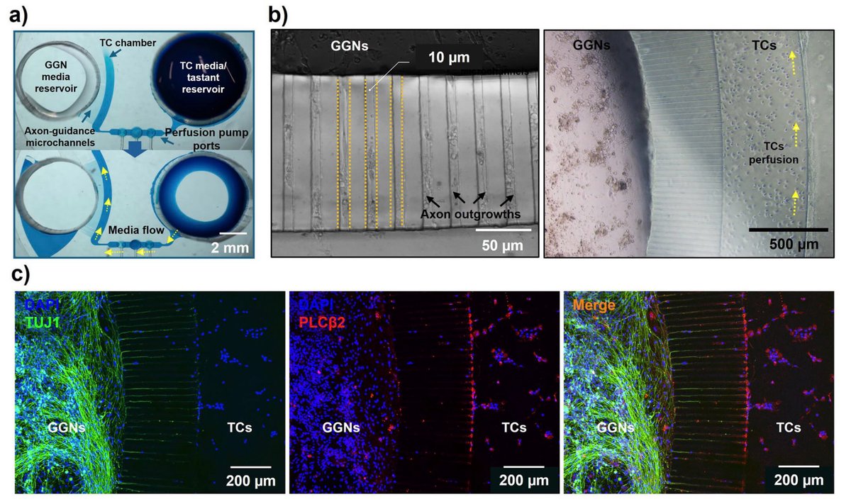
The new device addresses that question by placing primary taste cells and geniculate ganglion neurons in separate chambers. Tiny connecting channels allow neuronal axons, the long projections that transmit nerve signals, to grow toward the taste-cell chamber while limiting bulk fluid movement between the compartments.
A Pump Built Into the Chip
The team made the multilayer device from PDMS, a flexible silicone material widely used in microfluidics, using photolithography and soft lithography. Its flow layer contains culture chambers, reservoirs, and 10-micrometer-wide channels that guide axon growth.
A second layer contains pneumatic valves and an on-chip peristaltic pump. Like gently squeezing a flexible tube in sequence to move liquid forward, the valve sequence drives controlled one-directional fluid flow without an external syringe pump.
This integrated pumping system delivered tastants specifically to the taste-cell compartment. Researchers first cultured neurons from neonatal rats so their axons could extend through the channels, then introduced primary taste cells into the neighboring chamber.
Watching Sweet and Bitter Messages Travel
The cells were loaded with Fluo-4 AM, a dye that fluoresces when calcium levels rise. Because calcium changes accompany cellular activation, live fluorescence imaging let the team monitor signaling while exposing taste cells to sucrose, a sweet stimulus, or denatonium benzoate, a bitter stimulus.
For both stimuli, calcium activity appeared in taste cells first and subsequently in connected neurons. Neurons grown without taste cells showed little response when exposed to the same tastants, supporting the conclusion that the neuronal activity in the co-culture arose mainly from upstream taste-cell signaling.
The platform also let researchers wash away a stimulus and apply controlled repeat stimulation without disrupting the compartment boundary. Sweet and bitter compounds generated somewhat different calcium-response patterns, suggesting the setup can capture differences in signaling dynamics rather than simply detect whether cells respond.
Why This Matters
The device creates a more tissue-like experimental arrangement than a shared culture dish. It combines physical separation, directed fluid flow, axonal connectivity, and real-time calcium imaging in one platform, giving researchers tighter control over where a chemical arrives and when a cell responds.
That control could make the system useful for studying other taste modalities, taste dysfunction, and compounds that modify taste perception. Future work will need to show how well the platform handles additional cell types and whether its findings translate beyond this neonatal rat cell model.
Topics: Microfluidics · Brain & neurological health
Start here: Microfluidics explained