Source: TMOS, written by SAE Media Group (October 9, 2026). AI-generated summary by biochip.com, published . Not independently reviewed.
Key takeaways
- TMOS researchers built a nonlocal metasurface that performs quantitative phase microscopy without moving optical components.
- The device uses two wavelength-specific nanostructure sets to capture perpendicular phase measurements and reduce image noise.
- The system still requires electron-beam fabrication and precisely tunable illumination, limiting near-term deployment.
Researchers at the Australian Research Council Centre of Excellence for Transformative Meta-Optical Systems (TMOS) have built a multifunctional metasurface that performs quantitative phase microscopy in a compact optical component. Led by Professor Ann Roberts and TMOS PhD student Haiwei Wang, the work combines two wavelength-specific measurements that conventional systems typically collect separately. The approach could help shrink advanced microscopes into point-of-care devices for examining cells and tissues without staining them. That matters because many diseases, including cancers and sickle cell disease, change cells' shape, growth, or internal optical properties.
Making Nearly Transparent Samples Visible
Many biological samples are difficult to see under an ordinary microscope because they transmit light rather than strongly absorbing or reflecting it. Phase microscopy solves this problem by measuring small shifts in light waves after they pass through a specimen, rather than simply measuring brightness.
A useful analogy is looking at ripples moving across a pond. Even if the water itself is clear, an object beneath the surface changes the timing and pattern of the ripples. Likewise, cells alter the phase of transmitted light, creating information that can reveal their structures without dyes that may take time to apply or alter the sample.
From Phase Images to Quantitative Measurements
The TMOS device is designed for quantitative phase microscopy, or QPM. Unlike an image that only outlines a cell, QPM measures the exact phase shift across different regions of a sample.
Those measurements can be used to calculate properties including refractive index, which describes how much a material bends light, and the dry weight of tissue. Such values can provide objective information for biological research and diagnostic imaging, rather than relying only on a visual assessment of cell appearance.
Two Optical Jobs on One Surface
Metasurfaces are ultrathin materials patterned with structures about the size of light wavelengths. By arranging these nanoscale features precisely, engineers can make a flat surface control light in ways that conventional lenses and natural materials cannot.
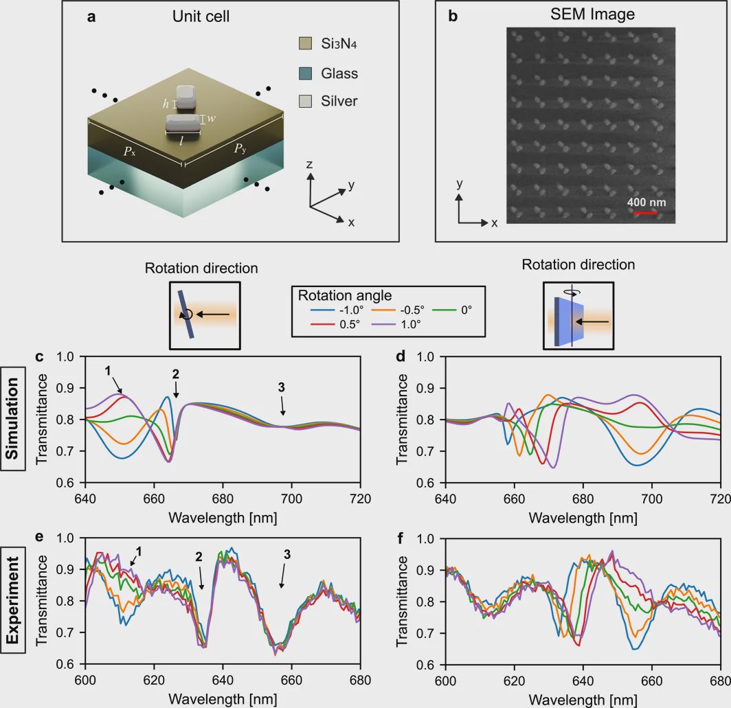
The team used a nonlocal metasurface, meaning it processes the image as a whole rather than redirecting each light ray independently like a standard lens. This makes it possible to position the component directly in front of the sample, avoiding the large gaps and multiple bulky elements common in microscope setups.
Conventional QPM generally needs phase measurements along two perpendicular directions. That can mean capturing two images and carefully realigning them, or completing a longer characterization process. The TMOS design incorporates both measurements into one surface, without the two signals interfering with each other.
Wavelength Multiplexing Reduces Noise
The researchers engineered separate sets of nanostructures to respond to different narrow bands of light. One set responds at one wavelength and the other at a different wavelength, allowing the metasurface to gather the needed optical information through wavelength multiplexing.
This arrangement also incorporates differential phase contrast, an imaging method that compares light changes across a sample to make subtle features clearer and reduce noise. The resulting setup can achieve quantitative phase microscopy without mechanically moving components.
“Here we are applying them to access different functionalities, which hasn’t been done before,” Wang said. The work appeared in Nanophotonics under the title “Wavelength and Polarization Multiplexed Nonlocal Metasurface for Quantitative Phase Microscopy.”
Why This Matters
A smaller, simpler QPM system could bring detailed cell imaging closer to clinics and other settings where a full microscope installation is impractical. TMOS identified potential uses in conditions where clinicians or researchers need to observe cell shape and growth patterns, including cancers, sickle cell disease, and neurodegenerative diseases.
The technology is not yet a finished point-of-care diagnostic product. The current implementation depends on electron-beam fabrication and precisely tunable illumination, both of which present practical barriers for lower-cost deployment. Commercial nanoimprint lithography may offer a more scalable manufacturing route.
Wang is now integrating the design into a commercial microscope using a simpler infrared light-emitting diode configuration. Longer term, the team aims to move toward miniaturized sensors that could support applications such as endoscopic imaging inside the body and wavefront sensing, which measures distortions in light.
Topics: Diagnostics & precision medicine
Start here: What is a biochip?