Source: Biosensors, by Zeeshan; Naeem Iqbal; Jaeyoung Choi (September 21, 2026). AI-generated summary by biochip.com, published . Not independently reviewed.
Key takeaways
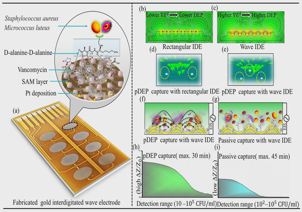
- A wave-shaped interdigitated electrode achieved 19% simulated bacterial capture, versus 12% for a rectangular electrode.
- The Pt-black-modified sensor detected S. aureus and M. luteus down to 10 CFU/mL within 30 minutes in 0.1× PBS.
- In skim milk, positive dielectrophoresis detected 100 to 100,000 CFU/mL, not the 10 CFU/mL sensitivity reported in buffer.
Researchers at Gachon University and Queen's University Belfast have built an electrochemical biosensor that uses an electric field to pull Gram-positive bacteria toward its sensing surface. The device combines a wave-shaped interdigitated electrode, computer-guided design, and a vancomycin capture layer to detect Staphylococcus aureus and Micrococcus luteus. In phosphate-buffered saline, the system detected bacteria down to 10 colony-forming units per milliliter within 30 minutes. The work shows how reshaping an electrode can improve the practical capture step that often limits rapid bacterial sensing.
Using Electric Fields to Gather Bacteria
The sensor relies on dielectrophoresis, the movement of particles in a nonuniform electric field. A simple analogy is debris being drawn toward the strongest part of a whirlpool. Here, an alternating electrical signal creates high-field regions that attract polarizable bacterial cells, concentrating them where the sensor can measure them.
This process is called positive dielectrophoresis when cells move toward stronger electric fields. Rather than waiting for bacteria in a liquid droplet to drift to the electrode by chance, the device actively guides them to the sensing interface. That concentration step can make a sparse bacterial sample easier to detect quickly.
A Wave Pattern Beats a Conventional Layout
Zeeshan, Naeem Iqbal, and Jaeyoung Choi compared conventional rectangular interdigitated electrodes with a wave-shaped version. Interdigitated electrodes are comb-like arrays of interlocking metal traces that generate electric-field gradients in the gaps between them.
The team used finite-element modeling, a computational method that breaks a complex physical system into small pieces for calculation, to model electric fields, dielectrophoretic forces, and bacterial transport. The simulations indicated that the wave-shaped interdigitated electrode created broader high-field regions and a larger effective capture area than the rectangular design.
Under positive dielectrophoresis conditions, the model predicted a bacterial capture efficiency of 19% for the wave-shaped electrode, compared with 12% for the rectangular electrode. The researchers then fabricated the wave-shaped design on a printed circuit board substrate, turning the modeled geometry into a compact sensing platform.
Turning Capture Into an Electrical Readout
The fabricated electrode was modified with platinum black, a rough platinum coating that increases the electroactive surface area. More surface area provides more locations for electrical interactions at the electrode-liquid boundary, strengthening the impedance signal used for detection.
The device used vancomycin as its capture probe. Vancomycin can bind structural features associated with Gram-positive bacterial cell walls, allowing the surface to recognize the target cells after the electric field brings them close. A custom-built 16-channel portable potentiostat performed sequential impedance measurements, tracking changes in electrical resistance and capacitance caused by bacteria binding at the surface.
With positive dielectrophoresis switched on, the wave-shaped sensor detected both S. aureus and M. luteus across a 10 to 100,000 colony-forming-units-per-milliliter range in 0.1× phosphate-buffered saline. Passive operation, without electric-field enrichment, had a narrower range of 100 to 100,000 colony-forming units per milliliter.
Performance in Milk
Food and clinical samples are usually more complicated than a laboratory buffer. In skim milk, positive dielectrophoresis supported a linear detection range of 100 to 100,000 colony-forming units per milliliter, while passive detection ranged from 1,000 to 100,000 colony-forming units per milliliter.
The shift in detection range illustrates a central challenge for biosensors: proteins, fats, salts, and other material in a real sample can alter how cells move and how surfaces respond. Even so, electric-field-assisted enrichment retained an advantage over passive capture in skim milk.
Why This Matters
Rapid bacterial testing depends on two linked tasks: finding rare cells in a sample and converting their presence into a reliable signal. This platform addresses both by using electrode geometry to concentrate cells and impedance measurement to read out capture without requiring culture growth as part of the reported 30-minute detection step.
The direct comparison between electric-field-assisted and passive operation also helps isolate the value of dielectrophoretic enrichment. Future work will need to establish how the approach performs across more complex samples and bacterial targets, but the study provides a design strategy for building electrochemical bacterial sensors around the physics of cell capture.
Topics: Biosensors · Infectious diseases
Start here: Biosensors explained