Source: RSC Advances, written by Zongjie Ma; Yan Guo; Wenjun Luo; Keyan Wei; Zhaobo Hu; Jiajun Huang; Yuting Chen; Chang-ye Hui (October 19, 2026). AI-generated summary by biochip.com, published . Not independently reviewed.
Key takeaways
- The engineered E. coli sensor uses indigoidine for lead and deoxyviolacein for mercury, producing distinct blue and purple signals.
- Reported detection limits were 6.74 nM for lead and 0.0631 nM for mercury, with dose-response patterns in spiked surface water and seawater.
- The study tested spiked water matrices, so it does not establish performance in naturally contaminated samples or routine field use.
Researchers have engineered a single strain of E. coli to produce different colors in response to lead and mercury in water. The whole-cell biosensor pairs two metal-responsive genetic circuits with two pigment pathways, so blue signals indicate lead and purple signals indicate mercury. Tests in spiked surface water and seawater preserved dose-response patterns, suggesting the approach could help with preliminary environmental screening. It is designed to make two contaminants easier to distinguish without relying on the costly instruments used in conventional metal analysis.
Two metal signals, one bacterial host
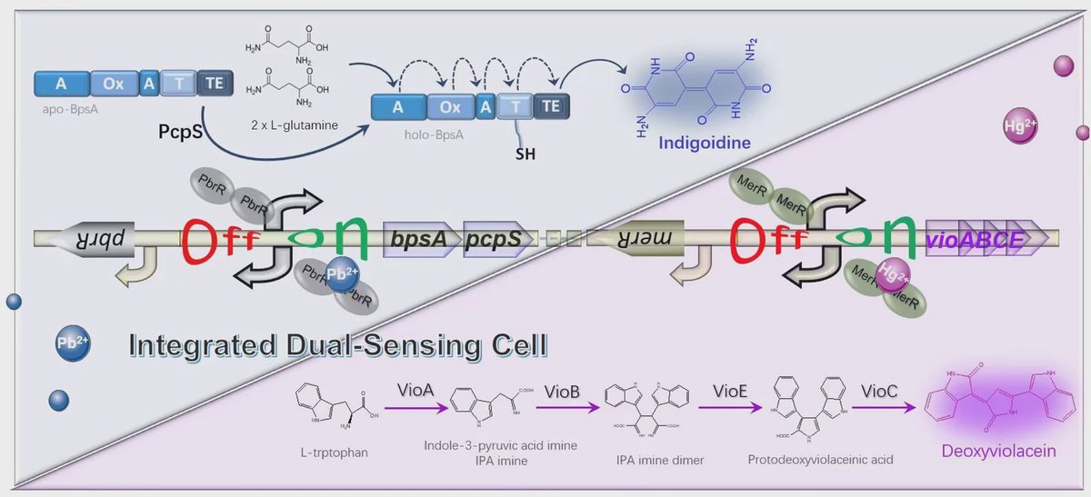
The team built the biosensor in E. coli TOP10, a laboratory strain, using one genetic module for each metal. Think of the modules as two separate alarms in the same cell: each responds to a different chemical trigger and switches on its own color-producing output.
Lead activates a regulator called PbrR, which drives production of indigoidine, a water-soluble blue pigment. Mercury activates a second regulator, MerR, which drives production of deoxyviolacein, a purple pigment. The two circuits sit in a single engineered construct, allowing the same bacterial culture to respond to both metals.
Separating the colors
The pigments also behave differently after the cells produce them. Indigoidine remains in the watery part of the sample, while deoxyviolacein can be collected in an organic solvent. That phase separation helps keep the signals from diluting one another, and the team measures them at different wavelengths: 600 nanometers for indigoidine and 570 nanometers for deoxyviolacein.
This design replaces an earlier pigment pairing that required a chemical oxidation step and did not separate the two readouts into distinct phases. The new combination avoids that oxidation step and creates a sharper blue-versus-purple contrast. The researchers say the colors can be distinguished by eye, while absorbance measurements provide a way to quantify each signal in a standard microplate format.
Detection performance and water tests
The reported detection limits were 6.74 nanomolar for lead and 0.0631 nanomolar for mercury. A detection limit describes the lowest concentration the test can detect under the study's conditions; it does not, by itself, show how reliably the device will perform in every water source. The researchers also report high selectivity against competing metal ions.
To assess performance in more complex samples, the team tested water matrices spiked with known amounts of the metals. The biosensor maintained clear dose-response relationships in both surface water and seawater. These tests suggest that other substances in those samples did not erase the color signals under the conditions tested.
Why This Matters
Laboratory techniques such as mass spectrometry can measure metals with high accuracy, but they require specialized equipment and trained operators. A living-cell sensor that produces visible colors could offer a simpler way to screen samples, particularly where access to those instruments is limited. Its ability to distinguish two metals in one culture is useful because lead and mercury can occur together in contaminated water.
The study establishes a proof of concept in spiked water samples, not a complete field-ready monitoring system. Further work would need to show how the sensor performs with naturally contaminated water and under practical sampling conditions. For now, the results point to a dual-color bacterial platform for preliminary screening, with the microplate measurements providing a quantitative check on the visual readout.
Topics: Biosensors · Environment & water
Start here: Biosensors explained
Related articles
- MXene-Polydopamine-Cyanobacteria Photobioelectrochemical Biosensor for Atrazine Detection in Environmental Waters
- Portable Biosensors Speed Food And Water Contamination Detection
- Electric-Field-Assisted Enrichment and Electrochemical Detection of Gram-Positive Bacteria Using an FEM-Guided Interdigitated Electrode Biosensor
- A target-responsive and signal-amplified DNA hydrogel biosensor for visual and quantitative detection of aflatoxin B1