Source: Lawrence Livermore National Laboratory, by Patricia Brady. AI-generated summary by biochip.com, published . Not independently reviewed.
Key takeaways
- Lawrence Livermore National Laboratory built a microscope-slide-sized gut-on-a-chip with 3D printed intestinal crypt and villus structures.
- The GelMA-based microfluidic device was developed to study interactions between human intestinal tissue and microorganisms.
- The report does not establish results from human testing or demonstrate specific disease, drug, or microbiome findings.
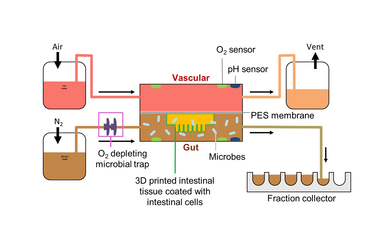
Lawrence Livermore National Laboratory researchers built a gut-on-a-chip that recreates key three-dimensional features of the human small intestine on a device about the size of a microscope slide. The microfluidic device, which guides fluids through tiny channels, is intended to give scientists a more realistic way to study interactions between intestinal tissue and microbes. Lindy Jang led the work reported in Biofabrication, while chip designer Michael Triplett and research engineer Rick Hynes contributed to the effort. The team used stereolithographic 3D printing, a method that hardens material layer by layer with light, to make a soft scaffold from gelatin-methacrylate, or GelMA, bioresins. Their design reproduces the intestine's crypt-villus architecture: cavities called crypts and finger-like villi that increase the organ's functional surface area. Existing organ-on-a-chip systems have helped researchers model disease and test drugs for more than 15 years, but many do not capture this much three-dimensional structure. By manufacturing the shape directly rather than waiting for cells to form it, the LLNL team sought to make a complex intestinal model more practical to create. The project was funded through the laboratory's Laboratory-Directed Research and Development Strategic Initiative.
A Tiny Model of a Vast Organ
The small intestine is a long, narrow tube that does an outsized share of the body's digestive work. Although it is only about 2 centimeters in diameter, the source describes it as extending 8 to 9 meters and containing roughly 250 square meters of surface area, comparable to a doubles tennis court.
That enormous surface is not packed into a smooth tube. It comes from a folded inner landscape filled with villi, the tiny projections that extend from the intestinal wall, and crypts, the recessed cavities between them. Together, these structures help create conditions that regulate cell behavior while expanding the area available for absorption.
Why Gut Structure Is Hard to Recreate
The human gut is also home to trillions of microorganisms, making it a living environment rather than simply a digestive pipe. The body contains roughly equal numbers of human and microbial cells, and the microbial community can influence human health. Studying that system directly is difficult because so many biological players and physical structures interact at once.
Organ-on-a-chip devices offer a scaled-down alternative. Think of them as a working miniature rather than a static model: living tissue is placed in a controlled device where researchers can manage the surrounding fluid and examine specific interactions. These in-vitro platforms, meaning systems studied outside a living organism, can support disease modeling and drug discovery while reducing reliance on animal testing.
Printing the Intestinal Landscape
Many chip-based organ models simplify the anatomy they represent. For the small intestine, that simplification can omit the three-dimensional crypt-villus pattern that gives the tissue much of its distinctive physical character. Hynes said that reproducing this architecture directly on a chip has been a persistent challenge.
One approach is to grow the structures from cells. But LLNL noted that this process can be time-consuming and have a high failure rate. The researchers instead drew on the laboratory's expertise in bioengineering and additive manufacturing to build the physical scaffold first.
The team used stereolithographic 3D printing, which uses light to shape a material into a designed form. Their printing material was a GelMA-based bioresin, a soft gelatin-derived material suited to making a tissue-supporting scaffold. They also used an enzyme called transglutaminase to crosslink proteins onto the scaffold, a chemical step that helps link protein components together.
Designed for Human-Microbe Studies
The resulting device combines the printed intestinal architecture with microfluidics, the controlled movement of very small volumes of liquid through channels. That combination is central to the model's purpose: creating a closer physical approximation of the small intestine while providing an experimental setting for human tissue and microbial interactions.
Jang described the gut as an exceptionally complex system and said the team developed a simpler research device intended to mimic it as closely as possible. The goal is not to shrink a full human intestine onto a slide. It is to preserve selected biological features that matter when researchers ask focused questions about intestinal tissue and its resident microorganisms.
Why This Matters
Structure affects biology. A flat layer of intestinal cells cannot fully stand in for tissue organized around cavities and projections, just as a flat street map cannot convey the elevation changes that shape how people move through a city. By incorporating crypts and villi into a fluidic chip, the LLNL platform addresses a physical feature that is often difficult to reproduce in laboratory models.
That matters for experiments involving the gut microbiome, the community of microorganisms associated with the digestive system. A more anatomically detailed model could help researchers examine how microbes and human intestinal tissue behave together under controlled conditions, without attempting to isolate every variable inside a living person.
What Comes Next
The LLNL work points toward organ-on-a-chip models that use fabrication techniques to add biological realism where it is most needed. As researchers continue to refine how living cells, soft materials, and flowing fluids work together on chips, models of the gut may become more useful for testing precise questions about one of the body's most complex environments.