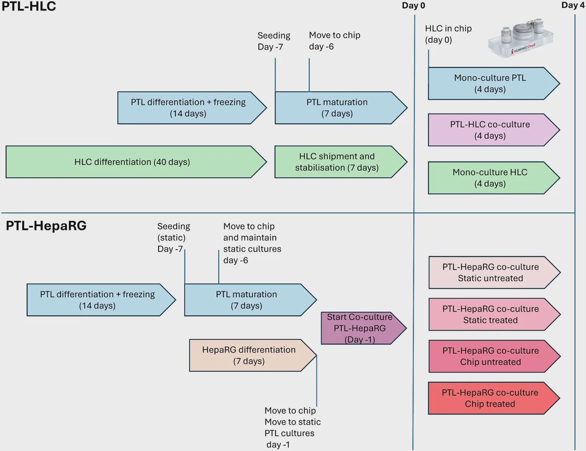
Source: Lab on a Chip, by Lenya de Brouwer; Victoria Pozo Garcia; Mostafa Kiamehr; Elisabeth Naderlinger; Tuğçe S Çobanoğlu; Beren Ataç Wagegg; Eva-Maria Dehne; Catherine Verfaillie; Sofia Moco; Paul Jennings; Anja Wilmes (September 29, 2026). AI-generated summary by biochip.com, published . Not independently reviewed.
Key takeaways
- Vrije Universiteit Amsterdam collaborators built a fully iPSC-derived kidney-liver organ-on-a-chip with liver organobodies and proximal tubule-like cells.
- A complementary PTL-HepaRG model converted ifosfamide into chloroacetaldehyde, demonstrating liver-mediated prodrug bioactivation.
- Chloroacetaldehyde concentrations were too low to cause adverse effects in renal tissue in the tested setup.
Researchers at Vrije Universiteit Amsterdam, KU Leuven, and TissUse GmbH have built a kidney-liver organ-on-a-chip using cells derived from human induced pluripotent stem cells, or iPSCs. The microfluidic system pairs liver-like cell clusters with kidney proximal tubule-like cells, allowing the tissues to communicate under flowing culture conditions. It produced distinct gene-expression changes in both tissues and supported the study of ifosfamide, a drug that must be metabolized by the liver before its effects can be assessed. The work offers a human-based platform for investigating how the liver and kidney jointly process drugs and respond to potentially toxic metabolites.
Connecting two organs under flow
The liver and kidneys work as a linked drug-processing system. The liver chemically modifies many drugs, while the kidneys filter compounds and metabolites from the blood for removal in urine. Studying either organ alone can miss how a liver-produced metabolite affects kidney tissue, much as testing one station in an assembly line cannot reveal what arrives from the station before it.
The team created an organ-on-a-chip system with hepatocyte-like cell, or HLC, organobodies and proximal tubular-like, or PTL, cells. HLC organobodies are three-dimensional clusters of liver-like cells, while PTL cells model the kidney tubules that help recover useful substances and secrete waste products. Microfluidic flow moved culture medium through the system, adding a physical feature that conventional static cell cultures lack.
A fully iPSC-derived model
iPSCs are adult cells reprogrammed into a stem-cell-like state, after which they can be directed to develop into different cell types. The kidney and liver components in this platform were derived from iPSCs, creating what the researchers describe as the first fully iPSC-derived kidney-liver organ-on-a-chip platform.
This approach could help researchers build models from a shared donor background. Because iPSCs can expand for long periods and be differentiated into several tissues, they may offer a more standardized and renewable source of human cells than primary tissue samples, which have limited availability and can lose specialized function in culture.
Co-culture changed both tissues
Growing the kidney and liver models together did more than place them in the same device. Gene set enrichment analysis, a method that looks for coordinated shifts across groups of genes, found distinct transcriptional responses in both cell types during co-culture.
In PTL cells, co-culture with HepaRG cells enriched pathways linked to signaling and cell polarity, the organized orientation that helps epithelial cells form functional barriers and transport surfaces. Co-culture with iPSC-derived HLCs enhanced gene programs related to metabolism and transport. In the HLCs, the presence of PTL cells affected intermediary metabolism, lipid processing, redox regulation, and plasma-protein synthesis.
Testing metabolism-dependent drug effects
The researchers also established a complementary system that combined PTL cells with HepaRG spheroids, three-dimensional clusters made from a human liver cell line with useful metabolic activity. They used this model to examine ifosfamide, a prodrug whose biologically active products arise after liver metabolism.
The PTL-HepaRG co-culture bioactivated ifosfamide into chloroacetaldehyde. That result demonstrates that the linked system can capture an important sequence: a liver-like tissue converts a compound, then a kidney-like tissue is exposed to the resulting metabolite. However, chloroacetaldehyde levels did not reach concentrations that caused adverse effects in the renal tissue under the conditions tested.
Why This Matters
Drug safety depends on more than whether a compound harms one isolated tissue. A medicine can be changed by liver enzymes, circulate as a new molecule, and then affect the kidney during clearance. A connected chip model could therefore help investigate toxicity and absorption, distribution, metabolism, and excretion processes in a setting that better reflects this organ-to-organ exchange.
The platform still needs further testing with compounds and exposure conditions that generate measurable renal injury. Its immediate value is as a controlled human in vitro system for asking how liver metabolism and kidney transport influence one another, and for refining models that may eventually support more informative preclinical drug testing.
Topics: Organ-on-a-chip
Start here: Organ-on-a-chip explained
Related articles
- Organ-on-a-chip platforms for disease modeling and in vitro diagnostic applications
- Organ-on-a-chip platforms for drug development, cellular toxicity assessment, and disease modeling
- Organ-on-a-chip technology replicates decades of human aging in just four days
- Researchers develop 'organ on a chip' for better drug testing