Source: Frontiers in Bioengineering and Biotechnology, by Po Hao; Jingrong Deng; Jian Xu (September 17, 2026). AI-generated summary by biochip.com, published . Not independently reviewed.
Key takeaways
- Hao, Deng, and Xu review organ-on-a-chip platforms that combine microfluidics, engineered tissues, and sensing for in vitro diagnostics.
- The review highlights real-time electrochemical, optical, fluorescent, and microelectrode-array sensing alongside disease modeling and biomarker screening.
- Clinical translation remains limited by physiological complexity, lack of standardization, and industrialization barriers.
A review in Frontiers in Bioengineering and Biotechnology examines how organ-on-a-chip systems could make in vitro diagnostics more biologically realistic. Po Hao, Jingrong Deng, and Jian Xu of Chongqing Three Gorges Medical College focus on devices that combine miniature fluid channels, engineered human tissues, and sensors to model disease and collect diagnostic signals. The central promise is straightforward: instead of examining cells in a static dish, researchers can observe tissues under controlled flow, mechanical forces, and continuous measurement. That could help narrow the gap between laboratory findings and human biology, where many candidate drugs and biomarkers fail.
Small Devices That Recreate Key Organ Functions
An organ-on-a-chip, often shortened to OoC, is a microfluidic device designed to reproduce selected features of an organ's environment. Microfluidics means controlling very small volumes of liquid in tiny channels, much like directing water through a miniature plumbing system. The technology combines this controlled flow with living cells and three-dimensional tissue structures.
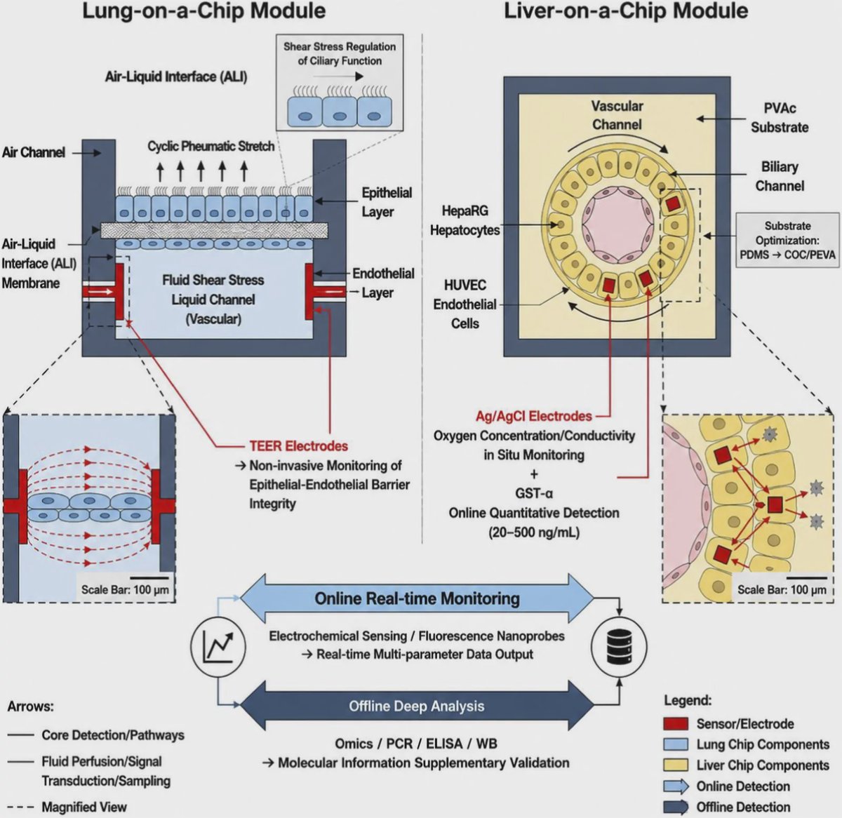
The review highlights a lung model as a classic example. A dual-channel alveolus-on-a-chip can place flowing culture medium in one channel to imitate blood circulation, while an air-liquid interface in another channel represents the lung's exposure to air. Cyclic stretching can then mimic breathing movements, allowing researchers to study the epithelial and endothelial tissue barrier under dynamic conditions.
Such systems aim to capture biological features that conventional two-dimensional cell cultures often miss, including tissue interfaces, fluid shear stress, and mechanical stimulation. The authors note that flow through vascular channels can promote synchronized beating of cilia, the tiny hair-like structures that help clear mucus in airways. A simplified airway chip using induced pluripotent stem cell-derived progenitor cells has also been used to model the effects of flow on ciliary development and to study conditions such as primary ciliary dyskinesia.
Why Sensors Matter for Diagnostics
The review frames organ-on-a-chip technology around in vitro diagnostics, or IVD, tests performed outside the body on biological samples or models. Traditional experiments often rely on endpoint measurements, meaning researchers collect a sample at the end and analyze it later. Chip platforms can instead integrate online sensing, which makes repeated or continuous measurements while an experiment is underway.
The detection options discussed include electrochemical biosensors, optical and fluorescent sensing, and microelectrode arrays. An electrochemical sensor converts a biological interaction into an electrical signal, similar in principle to how a smoke detector turns a physical change into an alarm. In an organ chip, these tools could track multiple signals in real time rather than relying solely on offline sampling and immunochemical tests.
This pairing of online monitoring and offline, in-depth analysis is important for applications such as disease modeling, biomarker screening, drug toxicity evaluation, and personalized diagnosis. A biomarker is a measurable biological signal linked to a condition or treatment response. The review argues that continuous, multi-parameter data could make chip models more useful for evaluating which biomarkers remain informative in a more human-like environment.
Organoids and Chips Solve Different Problems
The authors distinguish organ-on-a-chip systems from organoids, which are self-organizing three-dimensional cell structures derived from stem cells. Organoids can reproduce aspects of an organ's architecture and include multiple cell types, but they are commonly grown in relatively static conditions without controlled perfusion, mechanical forces, or integrated sensors.
Chips provide those controlled conditions, although they may use simpler cell arrangements. Combining the approaches creates organoids-on-a-chip, sometimes called chip-organoids. In a patient-derived organoid chip, patient-specific organoids are cultured in a microfluidic device for potential drug-sensitivity testing and biomarker discovery.
Materials and Multi-Organ Models
Materials are a practical obstacle as well as an engineering choice. The widely used silicone material polydimethylsiloxane, or PDMS, can absorb hydrophobic small molecules, potentially changing the concentration of drugs or other test compounds inside a device. The review identifies hydrogels such as GelMA, thermoplastics, and bio-based or degradable membranes as alternatives being explored for lower small-molecule adsorption and improved control over mechanical and chemical properties.
The field is also moving from single-organ devices toward multi-organ chips, which connect tissue models to study communication among organs. These systems could be valuable for examining pharmacokinetics and pharmacodynamics, terms that describe how a body handles a drug and how a drug affects the body. They may also help investigate how toxicity in one tissue influences another.
Why This Matters
Only about 10% of candidate drugs receive final approval, while 90% fail during clinical development, the review notes. No laboratory model can fully duplicate a person, but organ-on-a-chip platforms are intended to offer a closer approximation than static cultures or animal models for particular questions. Their value for diagnostics depends on whether the models can produce reliable, comparable signals that reflect clinically meaningful biology.
Important barriers remain. The review identifies insufficient physiological complexity, a lack of standardization, and challenges in clinical translation and industrialization. Artificial intelligence and machine learning may help interpret the large, multi-signal datasets produced by chips, but progress toward clinical IVD use will also require optimized models, standardized manufacturing and testing practices, and workable regulatory pathways.
The next stage is not simply making more sophisticated chips. It is determining which chip designs deliver reproducible measurements that clinicians, regulators, and diagnostic developers can use with confidence.
Topics: Microfluidics · Organ-on-a-chip · Diagnostics & precision medicine
Start here: Microfluidics explained
Related articles
- Microfluidic chips: recent advances, critical strategies in design, applications and future perspectives
- Organoids, Organ-on-a-Chip, and the Case for Online LC–MS
- Companion Diagnostics in Clinical Therapy: Current Applications and Future Directions
- A Comprehensive Review of Organ-on-a-Chip Technology and Its Applications