Source: Biomedicines, by Qiuyue Song; Fangrui Liu; Jinyan Zhao; Wenli Xie; Xianjun Fu; Kuo Xu (September 6, 2026). AI-generated summary by biochip.com, published . Not independently reviewed.
Key takeaways
- Shandong University researchers used tapered microfluidic channels to image zebrafish hearts without MS-222 anesthesia.
- Cardiac output measured 0.339 nL/s on-chip versus 0.279 nL/s under MS-222 anesthesia.
- The study reports short-term recovery only and does not establish long-term effects or performance across other disease models.
Researchers at Shandong University of Traditional Chinese Medicine have developed a microfluidic chip that holds zebrafish larvae still enough to image their beating hearts without anesthesia. The platform addresses a practical problem in heart failure experiments: the commonly used anesthetic tricaine methanesulfonate, also called MS-222, can suppress heart function and distort measurements. In zebrafish embryos treated with isoproterenol to induce heart failure, the chip produced higher cardiac measurements than anesthetized imaging, including cardiac output. The work suggests that hydrodynamic confinement, using fluid flow and channel shape rather than drugs or physical pressure, could support more physiologically faithful drug-screening assays.
Why Anesthesia Can Skew Heart Measurements
Zebrafish are widely used for phenotypic screening, a strategy that looks for visible or measurable changes in a whole living organism after treatment. Their transparent early-life stages make it possible to observe the heart directly, but conventional imaging usually requires anesthetizing the animals so they remain still.
That creates a complication when the measurement itself is cardiac performance. MS-222 can suppress cardiac activity, potentially making a weak heart appear weaker or masking the effect of a candidate treatment. The researchers designed their platform to keep larvae positioned for imaging while avoiding that pharmacological interference.
A Tapered Channel for Two Viewing Angles
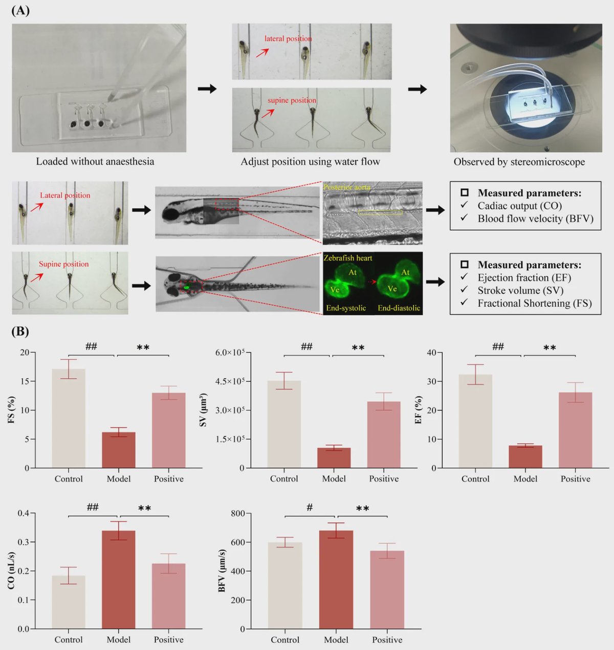
The microfluidic chip uses tapered immobilization channels to orient larvae for both lateral and supine imaging. Think of the channels as gently shaped holders: rather than clamping a larva in place, their geometry guides it into a consistent position while fluid-based confinement limits movement.
This orientation matters because different views support different measurements of heart structure and motion. The team collected cardiac output, blood-flow velocity, ejection fraction, stroke volume, and fractional shortening. Cardiac output is the volume of blood pumped per unit of time, while ejection fraction describes how much blood a heart chamber expels with each beat.
Testing a Zebrafish Heart Failure Model
The researchers exposed one-day-post-fertilization zebrafish embryos to 1 millimolar isoproterenol for 48 hours to create a heart failure model. Isoproterenol is a drug that stimulates beta-adrenergic receptors, placing stress on the cardiovascular system. A control group received embryo medium alone, while a positive-treatment group received isoproterenol plus 10 micromolar propranolol for the same 48-hour period.
The study compared measurements obtained with the chip's hydrodynamic confinement against measurements collected under MS-222 anesthesia. These were independent parallel experiments, meaning no individual larva was imaged by both methods.
Higher Cardiac Output Without MS-222
Cardiac parameters measured on the microfluidic platform were significantly higher than those measured under MS-222 anesthesia. The reported cardiac output was 0.339 ± 0.032 nanoliters per second with microfluidics, compared with 0.279 ± 0.023 nanoliters per second under anesthesia.
The device also achieved an immobilization success rate above 95%, with more than 90% of larvae correctly oriented for both lateral and supine imaging. The researchers reported that this was accomplished without physical compression or restriction of cardiac movement, two important design features when the goal is to observe natural heart mechanics.
Survival After Imaging
After one hour of continuous two-position photography, all larvae were released successfully from the chip and remained alive on visual inspection. In three independent experiments of 10 larvae each, the team observed no procedure-related mortality or morphological abnormalities during a 24-hour recovery period.
Those results indicate that the chip could be used repeatedly as part of a live-animal imaging workflow, although the reported observations cover short-term recovery rather than longer-term health or development.
Why This Matters
A heart assay must avoid changing the very physiology it is supposed to measure. By replacing anesthetic immobilization with a microfluidic design, this platform may help researchers distinguish disease-related changes in zebrafish cardiac function from changes caused by the imaging procedure itself.
The next useful step will be to test the approach across additional heart failure models and drug candidates, and to determine how well its measurements predict effects in other experimental systems. For zebrafish-based pharmacological screening, the study provides a practical route to collecting dual-angle heart data while larvae remain awake.
Topics: Microfluidics · Heart & vascular health
Start here: Microfluidics explained
Related articles
- Microfluidic Impedance Biosensor Chips Using Sensing Layers Based on DNA-Based Self-Assembled Monolayers for Label-Free Detection of Proteins
- Targeting solutions: Inside the ISEB’s Precision Diagnostics & Therapeutics research cluster at Case Western Reserve University
- What’s trending in point-of-care diagnostics?
- Quantum Nanosensors Could Reveal What Organs-on-Chips Miss