Source: Micromachines, written by Yunxing Lu; Xiaohan Wang; Qingchun Bai; Qiang Shi; Yiman Song; Lin Zhou; Zhenhua Wu; Hongju Mao (October 15, 2026). AI-generated summary by biochip.com, published . Not independently reviewed.
Key takeaways
- The integrated chip captures EVs, lyses them, generates droplets, and analyzes RNA mutations in one device.
- Tests using EVs from three cancer cell lines detected EGFR L858R and KRAS mutations, with an R-squared value of 0.9950 across sample inputs.
- The platform was tested with cell-line-derived EVs, and the study does not establish performance with patient samples.
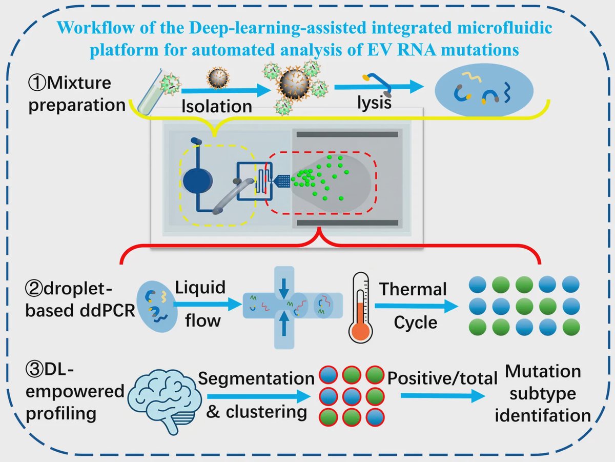
A research team in China has built a microfluidic chip that captures extracellular vesicles, releases their RNA, and analyzes mutation signals in droplets on one device. Deep-learning software automates the fluorescence-image readout, reducing the need for manual droplet counting and threshold adjustment. The team tested the system with vesicles from three cancer cell lines and detected EGFR L858R and KRAS mutations. The platform could help streamline analysis of vesicle RNA, although it has not yet been demonstrated with patient samples.
From vesicle capture to RNA analysis
Extracellular vesicles, or EVs, are tiny membrane-bound packages released by cells. They carry molecules that can reflect the state of the cells that produced them, including RNA with cancer-associated mutations. The researchers designed a chip to process EVs and their RNA without moving samples through a series of separate instruments.
The system uses magnetic beads coated with an antibody that binds CD63, a protein found on EVs. After the beads capture vesicles, the chip holds them in place while a lysis solution breaks the vesicles open and releases their contents. The sample then moves to the next stage for droplet formation and analysis.
Droplets create separate reaction units
The device uses reverse-transcription droplet digital polymerase chain reaction, or RT-ddPCR. In everyday terms, it divides a sample into many tiny reaction chambers, like sorting a mixed handful of objects into individual compartments. Reverse transcription converts RNA into DNA, and digital PCR detects target sequences in the resulting droplets.
This partitioning can help identify rare genetic targets that might be harder to distinguish in a single bulk reaction. The chip combines droplet generation with an array that holds droplets for analysis. The researchers report a linear response across a range of starting sample inputs, with an R-squared value of 0.9950, a measure of how closely the results tracked the input range.
Software reads fluorescence automatically
To interpret the droplets, the team combined Cellpose, a software tool for image segmentation, with K-means clustering, a method that groups data by similarity. The program identifies droplets in fluorescence images, removes background signal, measures gray-scale intensity, and classifies droplets as positive or negative without a person setting the threshold by hand.
The team evaluated the platform using EVs from three cancer cell lines: NCI-H1975 and SK-LU-1, which are lung cancer lines, and HepG2, a liver cancer line. The system detected EGFR L858R and KRAS mutations in these samples. These results demonstrate operation with cell-line-derived EVs, not clinical performance in people.
Why This Matters
Conventional EV RNA workflows often involve separate isolation, extraction, reaction setup, droplet generation, and readout steps. Each transfer can add hands-on work and create opportunities for sample loss or contamination. Bringing capture, lysis, droplet processing, and image analysis together addresses several of those workflow steps in one platform.
The study establishes a proof of concept for integrated EV RNA mutation analysis, with automated image processing as part of the system rather than a separate manual task. Testing with patient samples and establishing how the platform performs in clinical settings remain important next steps.
Topics: Microfluidics · Cancer
Start here: Microfluidics explained
Related articles
- Integrated “Lab-on-a-Chip” Microfluidic Systems for Isolation, Enrichment, and Analysis of Cancer Biomarkers
- Fabrication and Applications of Microfluidic Devices: A Review
- Recent Advances in Microfluidic Chip Technology for Laboratory Medicine: Innovations and Artificial Intelligence Integration
- Multifunctional microfluidic chip for cancer diagnosis and treatment