Source: Advanced Science, by Yaqing Lu; Yingyu Zhang; Xianwei Zhang; Mengxin Zhang; Fei Zhang; Jushan Sun; Yahui Yang; Kangbo Liu; Peng Wang; Shuying Luo; Wancun Zhang (September 27, 2026). AI-generated summary by biochip.com, published . Not independently reviewed.
Key takeaways
- UUU-DZ-tFNA detected uracil-DNA glycosylase with a reported detection limit of 0.025 mU/mL.
- A model combining exosomal UDG and clinical indicators reported 85.7% sensitivity, 100.0% specificity, and 92.8% accuracy for neuroblastoma risk stratification.
- The reported findings do not establish performance in broader independent clinical populations or routine care.
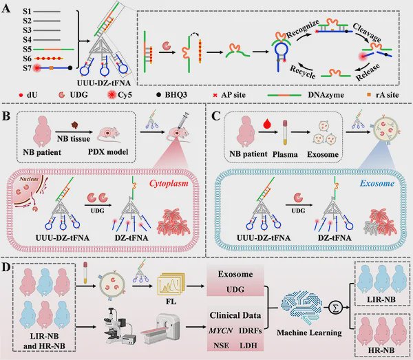
Researchers led by Yaqing Lu and colleagues at the Children's Hospital Affiliated to Zhengzhou University have built an engineered allosteric biosensor that detects uracil-DNA glycosylase, or UDG, inside neuroblastoma cells, animals, and patient-derived plasma exosomes. The device, called UUU-DZ-tFNA, measured UDG activity with a reported detection limit of 0.025 mU/mL and supported imaging-guided tumor removal in vivo. The team also combined exosomal UDG measurements with clinical indicators in a machine-learning model for neuroblastoma risk stratification. The work matters because neuroblastoma treatment decisions and emerging drug resistance can be difficult to assess without invasive tumor sampling.
A Sensor for a DNA Repair Enzyme
UDG is an enzyme that begins a DNA repair process known as base excision repair. Think of this pathway as a proofreading system: UDG identifies an inappropriate uracil base in DNA and removes it, allowing other enzymes to repair the damaged site. Cancer cells can exploit stronger DNA repair activity to survive damage caused by chemotherapy.
The researchers designed UUU-DZ-tFNA to recognize UDG directly rather than relying on the activity of another repair enzyme, apurinic/apyrimidinic endonuclease 1, or APE1. This distinction is important because many earlier in situ UDG methods generate signals that depend on both UDG and APE1, potentially complicating efforts to measure UDG itself.
How the Allosteric Design Works
The biosensor combines a DNA tetrahedral framework, a nanoscale structure built from DNA strands, with a DNAzyme, a DNA molecule engineered to perform an enzyme-like reaction. Its sequential activation design is intended to keep the signal quiet until UDG recognizes its target. Once activated, the system cycles its signal, amplifying the readout.
In laboratory testing, UUU-DZ-tFNA detected UDG at concentrations down to 0.025 mU/mL. The team reported that the platform was rapid, sensitive, and specific, and that it could visualize UDG at the cellular and animal levels in neuroblastoma models.
From Plasma Exosomes to Risk Stratification
Exosomes are tiny membrane-bound particles released by cells into body fluids, including blood. Because they can carry molecular information from tumors, they offer a potential route to assess disease without obtaining tumor tissue. The sensor measured UDG in plasma exosomes from patients with neuroblastoma.
The researchers incorporated exosomal UDG into a machine-learning model alongside MYCN amplification status, International Neuroblastoma Risk Factors, neuron-specific enolase, and lactate dehydrogenase. The reported model distinguished risk groups with 85.7% sensitivity, 100.0% specificity, and 92.8% accuracy.
Watching Drug Resistance
High-risk neuroblastoma often initially responds to chemotherapy but can later acquire multidrug resistance. The study links UDG monitoring to this problem because UDG helps initiate repair of DNA damage, including damage associated with chemotherapy exposure. The team reported that UUU-DZ-tFNA could monitor drug resistance in neuroblastoma.
The platform also enabled image-guided surgical excision in vivo. Instead of providing only a bulk measurement from disrupted tissue, in situ imaging aims to show where enzyme activity is occurring in living biological settings.
Why This Matters
Neuroblastoma is a highly variable childhood cancer: some tumors regress, while others spread rapidly and require intensive treatment. Current risk assessment often depends on tumor tissue, and biopsies can pose particular challenges for children with advanced disease. A blood-based measurement that captures tumor-associated enzyme activity could complement established clinical and molecular indicators.
The next step is to determine how this biosensor performs across broader clinical settings and during treatment decisions. If its measurements remain reliable beyond the reported study, direct UDG imaging and exosomal testing could help researchers study how DNA repair activity tracks with neuroblastoma risk and chemotherapy response.
Topics: Biosensors · Cancer
Start here: Biosensors explained
Related articles
- AI-Enhanced Photonic Biosensor Could Help Detect Glioblastoma Tissue
- Microfluidic biosensors for biomarker detection in body fluids: a key approach for early cancer diagnosis
- Liquid Biopsy in Breast Cancer: Biomarkers and Clinical Applications
- Companion Diagnostics in Clinical Therapy: Current Applications and Future Directions