Source: iScience, written by Zhiheng Yu; Heshuang Dong; Xiaolin Chen; Mengjiang Xu; Ren Liu; Jingjing Wang; Cunliang Yang; Ke Wang; Xianyuan Zhao; Hao Jia; Jijun Feng (October 12, 2026). AI-generated summary by biochip.com, published . Not independently reviewed.
Key takeaways
- The graphene oxide-coated microfiber detected ELANE at 3.57 × 10⁻²⁰ M and lactoferrin at 1.28 × 10⁻¹⁹ M.
- The sensor tracked distinct ELANE and lactoferrin release patterns in inflamed mice and showed different responses for septic patients and healthy controls.
- Clinical validation was preliminary, and the reported summary does not give the cohort size or establish improved patient outcomes.
Researchers at the University of Shanghai for Science and Technology and collaborating institutions developed a graphene oxide-coated optical microfiber sensor that detects two proteins released by activated neutrophils. The device uses DNA aptamers, short DNA molecules that bind specific targets, to recognize neutrophil elastase and lactoferrin. In tests, it detected both proteins at concentrations far below the reported sensitivity of conventional ELISA assays. The team also used it to track changing protein levels in a mouse inflammation model and to distinguish septic patients from healthy controls in a preliminary clinical cohort.
A fiber that reads protein binding
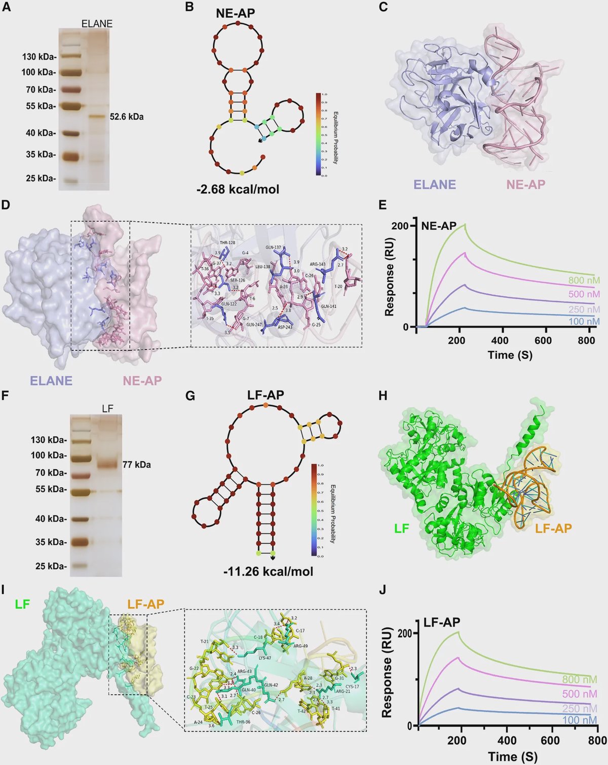
Think of the sensor as a tiny light guide with an optical fingerprint that changes when a target molecule attaches to its surface. The researchers coated an optical microfiber with graphene oxide, a thin carbon-based material, and attached aptamers designed to bind either neutrophil elastase, also called ELANE, or lactoferrin, or LF.
When a target protein binds to its matching aptamer, the change shifts the light spectrum toward longer wavelengths, a signal known as a red shift. The sensor reads that shift to detect the protein. The study says the probe can be immersed directly in a sample, and reports detection limits of 3.57 × 10⁻²⁰ M for ELANE and 1.28 × 10⁻¹⁹ M for LF.
Two proteins offer different timing clues
Neutrophils are immune cells that release stored proteins as they respond to infection or tissue injury. When that activity becomes excessive, the resulting inflammation can contribute to sepsis and tissue damage. Measuring neutrophil-related proteins may therefore help researchers follow how inflammation changes over time.
ELANE is stored in neutrophils’ primary granules and can be released within minutes to hours after activation, making it an early signal of inflammation. LF comes from secondary granules and tends to appear later and remain elevated longer. Tracking both could provide a fuller picture of neutrophil activity than measuring either protein alone.
Tests in mice and people
In mice given lipopolysaccharide to induce inflammation, the sensor detected different release patterns for ELANE and LF over time. Those differing patterns are consistent with the proteins’ distinct release timing and show how the device can follow changing marker levels in an experimental model.
In a preliminary clinical validation, the researchers reported different sensor responses in samples from septic patients and healthy controls. The study summary does not provide the cohort size or enough detail to assess how reliably the sensor would distinguish patients across a broader clinical population.
Why This Matters
The reported detection limits were eight orders of magnitude lower than those of ELISA, a widely used laboratory method for measuring proteins. That sensitivity could help researchers investigate low-level changes in neutrophil activity, including early signals that standard tests may miss. The device also uses an optical readout, which the researchers say avoids electromagnetic interference and could be miniaturized.
These findings establish a promising sensing platform, not a ready-to-use sepsis test. Larger clinical studies would be needed to determine how well it performs with real patient samples and whether it adds useful information to existing diagnosis. The next step is to test its accuracy and practicality in broader clinical settings.
Topics: Biosensors · Immunity & inflammation · Infectious diseases
Start here: Biosensors explained
Related articles
- PV-See: A novel Escherichia coli-based biosensor for PVC microplastic detection
- NEKfla: A Genetically Encoded Förster Resonance Energy Transfer Biosensor for Live-Cell Monitoring of Early NIMA-Related Kinase 7–NLR Family Pyrin Domain Containing 3 Inflammasome Signaling
- Electric-Field-Assisted Enrichment and Electrochemical Detection of Gram-Positive Bacteria Using an FEM-Guided Interdigitated Electrode Biosensor
- Interfacial Engineering of Biosensors for Bloodstream Infection Diagnosis