Source: Biotechnology Journal, written by Sarah Devriese; Flavio Bonanini; Anna F Bekebrede; Sara Shafiei; Dorota Kurek; Coen Govers; Hans Bouwmeester; A Paul Vos; Remko van Vught; Jaap Keijer; Vincent C J de Boer (November 1, 2026). AI-generated summary by biochip.com, published . Not independently reviewed.
Key takeaways
- A liquid-suspendable phosphorescent sensor measured oxygen consumption in microliter-scale gut-on-a-chip channels.
- The method tracked chemically induced metabolic changes in Caco-2 and colon organoid tubule models.
- Measurements are chamber-level and require static conditions, so the approach does not map oxygen across the tissue during flow.
Researchers at Wageningen University and Mimetas showed that a liquid-suspendable oxygen sensor can track changes in cell respiration inside a commercial gut-on-a-chip. The method works in microliter-scale channels without building a sensor into the device or inserting a probe. The team tested it with two gut models: Caco-2 cells and colon organoid-derived cells. That could make it easier to measure metabolism in chip models whose small, enclosed channels are difficult to access.
Reading oxygen in a small channel
Oxygen use offers a window into mitochondrial activity. Mitochondria use oxygen as part of the process that produces energy for cells, so changes in oxygen consumption can signal changes in cellular respiration.
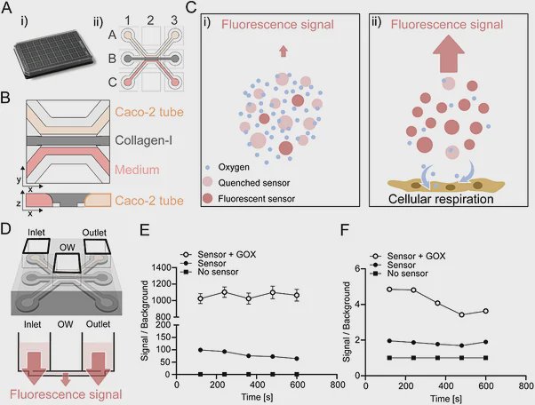
The sensor works a little like a glow that dims when oxygen is nearby. It is a cell-impermeable phosphorescent probe, meaning it stays outside cells and emits light after excitation; oxygen molecules quench that signal. By measuring the signal over time, researchers can track changes in oxygen around the cells.
Testing the approach in gut models
The team added the dissolved sensor to the culture medium in a commercially available microfluidic platform. They first checked whether the sensor signal could be detected in the device’s small channels, then developed a workflow to measure oxygen consumption in Caco-2 cell tubules and adult stem cell-derived colon organoid tubules.
To test whether the method could follow changing cell activity, the researchers added established mitochondrial respiration modulators in sequence. The study reports that time-resolved oxygen measurements were feasible in both ready-to-use gut-on-a-chip models and that chemically induced changes in oxygen metabolism could be monitored in real time.
What the sensor adds
Existing approaches to oxygen measurement in organ-on-a-chip systems can require a custom device with sensors built in, or a probe inserted through an access point. The liquid-suspendable method avoids those changes to the chip, and the researchers read its signal with a standard plate reader.
The approach does have a practical boundary: the reported sensor measures oxygen at the chamber level, not as a detailed map of oxygen across the tissue. The measurement also requires a static period, so flow through the chip must be interrupted while the signal is recorded.
Why This Matters
Organ-on-a-chip models aim to reproduce features of human tissues in compact devices, but their small size can make it hard to measure how cells function. Adding a dissolved sensor offers a way to assess respiration without redesigning the platform, which may help researchers study metabolic responses in more complex cell models.
The work establishes a measurement method in Caco-2 and colon organoid gut models. Further studies will need to show how it performs across other chip designs and biological applications.
Topics: Organ-on-a-chip · Diabetes & metabolism
Start here: Organ-on-a-chip explained