Source: iScience, written by Hongji Xu; Ruiqi Gu; Arthur Georg Serville Torvund; Iver Grønlien; Angelos Xomalis; Hai Son Nguyen; Zhengping Yu; Zhenyu Zhao (October 19, 2026). AI-generated summary by biochip.com, published . Not independently reviewed.
Key takeaways
- The metasurface measured q-BIC and EIT responses to screen colorectal tumor-associated fibroblast subtypes.
- MyCAFs shifted the q-BIC resonance from 0.92 to 0.86 THz, while apCAFs shifted it to 0.94 THz.
- The study does not establish the sensor’s performance in routine clinical samples.
Researchers have tested a terahertz biosensor that distinguishes two types of fibroblasts associated with colorectal tumors by reading how cells shift the device’s light response. The team, including researchers at Shanghai Normal University, Fudan University Shanghai Cancer Center, and the Norwegian University of Science and Technology, built the sensor to identify cell subtypes without labels. Its two sensing channels produced opposite frequency-shift patterns for pro-tumor and anti-tumor fibroblasts. The results suggest a possible way to screen these cells, though they do not establish clinical performance.
A sensor with two ways to read cells
The biosensor is a patterned surface, called a metasurface, made from tiny coupled split-ring structures. Like a finely tuned instrument that changes its note when something nearby alters its surroundings, the surface shifts its response when cells change the local electrical environment. The researchers measured those shifts using terahertz time-domain spectroscopy, which sends terahertz pulses at the surface and records how they pass through it.
The device supports two distinct optical responses. One is a quasi-bound state in the continuum, or q-BIC, a resonance that concentrates electromagnetic energy near the surface; the other is electromagnetically induced transparency, or EIT, a transmission feature created by interference between resonances. The team read the q-BIC with one polarization of terahertz light and the EIT response with the perpendicular polarization, giving the sensor two measurements from one platform.
Different fibroblasts, opposite shifts
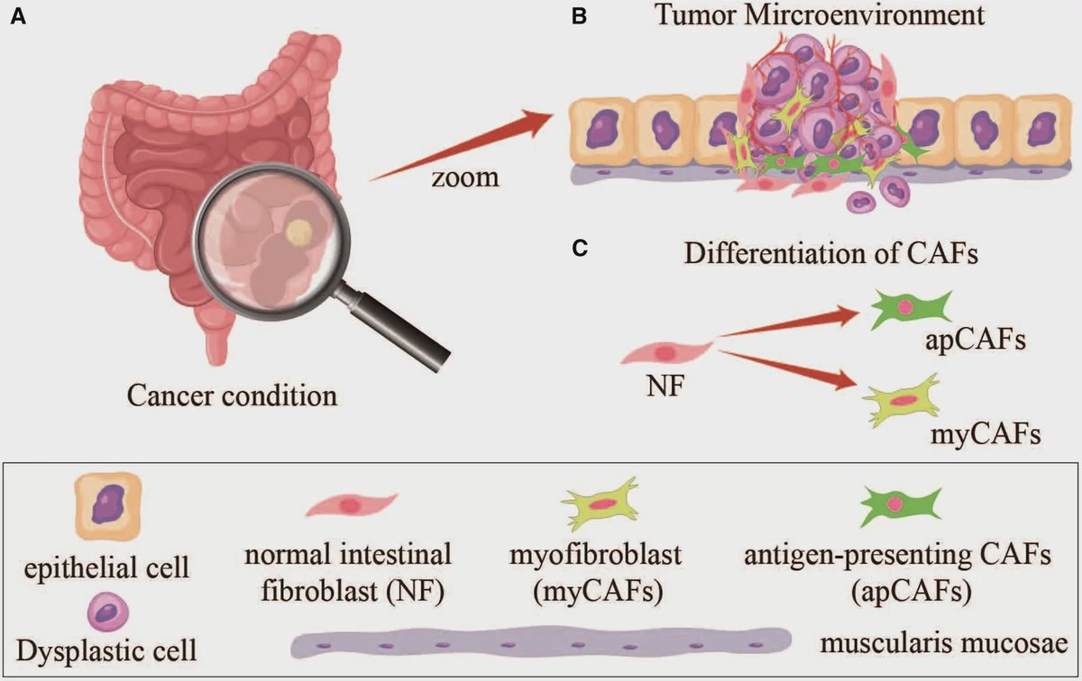
The researchers placed normal intestinal fibroblasts, myofibroblasts, and antigen-presenting cancer-associated fibroblasts on chemically prepared metasurfaces. Myofibroblasts, abbreviated myCAFs, are described in the study as pro-tumorigenic. Antigen-presenting CAFs, or apCAFs, are described as anti-tumorigenic. The team compared their responses with a control surface that had no cells attached.
For the q-BIC channel, the control resonance was at 0.92 terahertz. Normal fibroblasts shifted it slightly lower, to 0.91 terahertz, while myCAFs produced a larger redshift to 0.86 terahertz. ApCAFs shifted the resonance higher, to 0.94 terahertz. The EIT channel showed the same overall direction: myCAFs shifted its resonance lower, and apCAFs shifted it higher, although the reported summary does not give numerical EIT shifts.
The researchers attribute these changes to differences in the cells’ surface markers and the resulting local dielectric environment, meaning how the material around the sensor responds to an electric field. Flow cytometry and gene-expression analyses supported the presence of distinct markers associated with the fibroblast subtypes. Those tests provide biological context for the terahertz readout, but the reported findings do not show that the sensor can classify tumors in routine clinical samples.
Why This Matters
Fibroblasts in a tumor can have different effects, so identifying their subtype may help researchers understand the tumor environment. Current approaches such as flow cytometry and gene-expression profiling can be time-consuming and costly, the study notes. A label-free sensor could offer a different way to screen cells, using their effect on a surface resonance rather than adding a labeling reagent.
The work demonstrates a dual-channel sensing approach with distinct responses to the tested cell types. The next important question is whether those patterns remain reliable across larger sets of patient-derived samples and under conditions relevant to clinical testing. The study establishes a promising laboratory measurement, not a validated diagnostic test.
Topics: Biosensors · Cancer
Start here: Biosensors explained
Related articles
- AI-Enhanced Photonic Biosensor Could Help Detect Glioblastoma Tissue
- Tracking CDK1 Dynamics With a Novel Biosensor Mouse and Clinical Cohorts to Predict CDK4/6 Inhibitor Sensitivity in ER+ Breast Cancer
- Engineered Allosteric Biosensor for In Situ DNA Glycosylase Detection in Neuroblastoma Risk Stratification and Drug Resistance Monitoring
- Circulating Tumor Cells: Overcoming Detection Challenges